Synonym(s)
HistoryThis section has been translated automatically.
DefinitionThis section has been translated automatically.
Localized infectious skin disease caused by leishmania, which develops after the bite of an infected mosquito (sand flies -Phlebotomus papatasi) and is characterized by phased clinical symptoms.
You might also be interested in

PathogenThis section has been translated automatically.
Leishmania spp; see Table
Occurrence/EpidemiologyThis section has been translated automatically.
The incidence rates worldwide are 0.7-1.2 million per year.
90% of the diseases occur in the "Old World" (where the olive tree grows): Mediterranean regions, Africa (Algeria, Kenya, North Sudan, Ethiopia) Asia (Afghanistan, Iran, Syria), 10% in the "New World" (Texas, Brazil, Mexico, Bolivia, Peru).
The Leishmania species L. major, L. tropica, L. aethiopica, L. infantum (L. infantum = typical pathogen of leishmaniasis in Mallorca) mainly cause the diseases of the "Old World".
L. mexicana and L. brasiliensis cause the leishmaniasis of the "New World".
In non-endemic countries, the infections affect travelers or immigrants.
Humans are an accidental host. Wild rodents, especially rabbits, serve as a natural reservoir of leishmania.
In Afghanistan, gerbils are the natural reservoir in the desert-like regions of northern Afghanistan. The sand flies are also found in the burrows of these rodents, so that a purely zoonotic transmission cycle exists here. Humans are only accidental hosts.
In the Kabul area, more than 200,000 people have contracted leishmaniasis (Leishmania tropica. The infected person is the main reservoir. Sand flies are also vectors.
EtiopathogenesisThis section has been translated automatically.
Cutaneous leishmaniasis is transmitted in the "old world" by the female sand or butterfly mosquito of the genus Phlebotomus; in the "new world" by Lutzomyia. Promastigotes are transmitted into the skin during the biting process.
ManifestationThis section has been translated automatically.
Age at first manifestation:
- 5-9 years: 30% of infected persons
- < 20 years: 70% of infected persons
LocalizationThis section has been translated automatically.
Predilection sites are the uncovered skin areas:
- face and neck: 60%.
- upper extremity: 35%
- lower extremity: 10%.
ClinicThis section has been translated automatically.
After the bite of the sand fly, first an erythema and then a papule appear.
A distinction is made between:
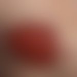
- Nodular form (regular picture): Incubation period 2-3 weeks up to 1 year. After the bite of the sand fly, first an erythema forms and then a 0.1-0.3 cm large, reddish-brown, succulent, slowly growing, itchy red papule. Regional lymphadenitis possible. No general symptoms. Within weeks, gradual but steady growth into a red-brown, painful (bright, cutting but not throbbing pain) nodule/node with a tightly stretched surface. Multiple nodules are rarer. Lymphangitis only complicated by bacterial overlay. The majority of nodules/nodes heal spontaneously without treatment after a period of several months with scarring. In infections caused by Leishmania tropica, pronounced local inflammatory reactions with marked swelling and erythema are observed.
- Ulcerative form: Development from the nodular form. Soft, encrusted ulcerations that usually heal spontaneously after a year (annual bumps, self-limiting). Formation of prominent scars.
- Recurrent form (recurrent cutaneous leishmaniasis - see below Leishmaniasis cutaneous recurrent leishmaniasis).
- Chronic hyperergic cutaneous leishmaniasis: This particular form is divided into a persistent and a recurrent form. It is characterized by reddish-brownish, lupoid plaques reminiscent of sarcoidosis or lupus vulgaris.
- Diffuse (anergic) cutaneous leishmaniasis: multiple, non-ulcerating papules and nodules that can spread symmetrically over the face and trunk as the disease progresses. Immunologically, there is a selective anergy of the immune system against Leishmania with high parasite density in the lesions. Triggers are L. amazonensis, L. mexicana, L. guyanensis, L. aethiopica. In Ethiopia and Sudan, this form occurs in about 20% of patients.
- Mucocutaneous leishmaniasis: severe form of initial cutaneous leishmaniasis (Espundia). It develops from originally cutaneous foci that spread to the mucous membrane area.
HistologyThis section has been translated automatically.
Acanthotic, in places pseudoepitheliomatous surface epithelium with parakeratotic serum crust. Dense, diffuse, granulomatous infiltrate of neutrophil granulocytes, plasma cells, lymphocytes, epitheloid-cell histiocytes and multinucleated giant cells in varying densities permeating the entire dermis. Numerous intracellular pathogens are found in fresh forms, particularly in the Giemsa stain. Basically, a granulomatous pattern or a pseudolymphatic pattern can be detected.
Immunohistologically, (indirect) detection by means of CD1a expression is successful in a higher percentage of cases.
Detection of the Leishmania antigen using PCR (also possible on formalin-fixed material). Species differentiation (PCR) is possible but time-consuming.
In older foci, the granulomatous component with small, isolated or confluent, epithelioid cell nodules predominates (DD: tuberculosis cutis luposa). Pathogen detection is then very sparse or even absent.
Differential diagnosisThis section has been translated automatically.
The initial clinical diagnosis of "cutaneous leishmaniasis" is only correctly made in around 1/3 of patients (in non-endemic countries). The initial diagnoses are mainly tumors (35%), infections of other origin (7%), insect bites and ulcers (10%) (Guillet C et al.2024). In this respect, the (clinical and histological) differentiation from other diseases is important:
Clinical:
- Boils (highly acute event, painfulness, fluctuation)
- Basal cell carcinoma (lack of inflammatory component)
- Eosinophilic granuloma (brown-red; follicular accentuation of the surface; histology is diagnostic)
- Ecthyma (highly acute, ulcerated with sharp edges; localization mostly lower extremity)
- Frambösia (country of origin)
- Syphilitic primary effect (localization)
- lupoid rosacea (age of patient; mostly disseminated follicular papules and pustules; rosacea face)
- Tuberculosis cutis luposa (few raised plaques and atrophy; rather brown-red, never strong red; self-infiltrate).
- Psoriasis (no history of travel, mostly disseminated, Auspitz phenomenon positive)
Histologically:
- Cutaneous B-cell pseudolymphoma (detection of germinal centers, which are absent in leishmaniasis)
- Lupus vulgaris (important in cases of long-term leishmaniasis, as it is often difficult to detect the pathogen; accompanying plasma cell reaction tends to indicate tuberculosis)
- Histoplasmosis (granulomatous infiltrate; detection of spores in the PAS preparation).
General therapyThis section has been translated automatically.
The pathogens respond to different treatments: While local measures are often sufficient in the case of an infection caused by Leishmania species from Europe and Africa ("Old World"), systemic therapy is the first choice for cutaneous and mucocutaneous forms of the "New World".
In the case of individual lesions in cosmetically undisturbed localisation, spontaneous healing can be awaited (poor scar formation). Excision or cryosurgery are no longer recommended, as pathogens are removed from the lesion and thus the development of immunity is made more difficult. There is a risk of disseminating the pathogens that are already present in the lymph channels.
External therapyThis section has been translated automatically.
An effective antibiotic substance against L. major that has been proven in several studies is a 15% paromomycin ointment in 12% methylbenzethonium chloride (available as Leshcutan/Teva from international pharmacies; licensed in Israel; have it prepared in Vaselinum album for magistral prescriptions ). Applications 2 times/day over a period of 10-14 days. If necessary, repeat the therapy cycle after 14 days (topical paromomycin is the chosen first-line therapy in around 40% of cases).
Local infiltration with antimony preparations: 1-3 ml meglumine antimonate (e.g. Glucantim) = 85 mg Sb/ml (depending on the size of the lesion; with or without the addition of triamcinolone acetonide); initially carry out infiltration mainly in the peripheral area, as this is where the highest pathogen density is found. Later also inject in the center of the lesion. Carry out the procedure 5 days a week; period: 4-5 weeks; usually quite painful procedure.
Alternative (or complementary): Infrared therapy ( hyperthermia): heat to approx. 55 °C for 5 minutes, repeat after 3 weeks if necessary (death of the pathogens; immune response is possible).
Alternative: Cryotherapy (liquid nitrogen in spray or contact method).
Alternative: Success with photodynamic therapy (PDT) has been reported in individual cases (2 irradiations per week over 4 weeks (Metvix; Waldmann PDT 1200 L, 100 J/cm2).
Internal therapyThis section has been translated automatically.
Antimony preparations (antimony = stibium = Sb): Generally the drug of choice for infections with Leishmania brasiliensis! Sodium stibogluconate (e.g. Pentostam) contains 10% stibium (100 mg/ml); meglumine antimonate (e.g. Glucantime) contains 8.5% stibium (85 mg/ml). Dosage: 20 mg/kg bw/day i.m. (painful) or slowly (10-20 min.) via small caliber needle i.v. Caution! Venous thrombosis! When administered i.v., the drug should be dissolved in 50 ml 5% glucose. Duration of therapy: 20 days for cutaneous forms and 28-30 days for mucocutaneous forms. If toxic side effects occur, reduce dose by 2 mg/kg bw Sb. Clinical healing often only occurs 4-6 weeks after the end of treatment. Individual uncomplicated leishmaniasis lesions can also be treated intralesionally (apply 1-3 ml of the undiluted solution from the edge into the lesion! Carry out the procedure 1-2 times/week. Duration of treatment: 3-6 weeks, depending on how acute the lesion is. Caution! Painfulness, toxicity for heart and liver!
Pentamidine diisethionate (e.g. Pentacarinat®): Therapy of choice for diffuse cutaneous leishmaniasis and for treatment failures of mucocutaneous forms. Dosage: 4 mg/kg bw/day i.m. once/week for at least 4 months. Recent studies have also shown efficacy in cutaneous leishmaniasis at a low dosage of 2 mg/kg i.m. every 2nd day up to a total of 7 injections.
Liposomal amphotericin B (e.g. Ambisome®): Drug of 2nd choice if pentavalent antimony is contraindicated or ineffective. ED: 0.5-1.0 mg/kg bw, every 2nd day, 20 doses in total.
Ketoconazole (e.g. Nizoral®): Drug of choice for infection by Leishmania mexicana, Leishmania panamensis and Leishmania major. Not or insufficiently effective against Leishmania brasiliensis, Leishmania tropica and Leishmania aethiopica. Dosage: 600 mg/day (evening) for 4 weeks.
Fluconazole (e.g. Diflucan®): Very effective in studies for infection with L. major. Dosage: 200 mg p.o. once/day for 6 weeks
Miltefosine (Impavido®): Adults and children from 3 years of age: 1.5-2.5 mg/kg bw/day for 28 days. Max. TD: 150 mg. Immunocompromised patients ( HIV-infected) may require longer treatment. The therapeutic successes in cutaneous leishmaniasis of the New World are also promising. Approval for this indication has been applied for in Pakistan and Colombia. Recent reports indicate that miltefosine can also be used successfully in cutaneous L. of the Old World.
Progression/forecastThis section has been translated automatically.
The course is usually without complications.
Rarely (!) accompanying lymphangitis, erysipelas or pyoderma.
Spontaneous healing of scars after about one year in the majority of infected persons
The disease leaves behind an immunity against the triggering species for several years.
ProphylaxisThis section has been translated automatically.
Insecticides, suitable clothing, mosquito net to avoid mosquito bites. If necessary, removal of reservoirs (dogs, rodents).
Vector control is also important. Phlebotomus papatasi the sand fly has limited flight characteristics with a daily radius of action of only 50 m and a maximum flight altitude of 1.50m. Further prevention measures can be based on these characteristics.
TablesThis section has been translated automatically.
Species |
Application |
Therapy |
Application/daily dose |
Therapy duration |
L. major |
external |
paromomycin 15%. |
2 times/day |
10 days, possible repetition |
Alternative: Heat treatment e.g. with infrared lamp (PDT) |
Heat up to 55 °C for 5 minutes |
1-2 times |
||
Alternative: Meglumin antimonate (85 mg stibium/ml) |
1-3 ml local infiltration |
1-2 times |
||
systemic |
Ketoconazole |
600 milligrams p.o. |
over 4 weeks |
|
Alternative: Antimony preparation |
20 mg/kg bw Sb-Ä. i.m. or i.v. |
cutaneous: 20 days |
||
mucocutaneous: 28 days | ||||
| ||||
L. tropica |
external |
Meglumin antimonate (85 mg stibium/ml) |
1-3 ml local infiltration |
1-2 times |
systemic |
Antimony preparation |
20 mg/kg bw Sb-Ä. i.m. or i.v. |
cutaneous: 20 days |
|
mucocutaneous: 28 days | ||||
| ||||
L. aethiopica |
external |
insufficient data situation |
||
systemic |
Pentamidine |
once/week 4 mg/kg bw |
over 4 months |
|
| ||||
L. brasiliensis |
external |
not indexed |
||
systemic |
Antimony preparation |
20 mg/kg bw Sb-Ä. i.m. or i.v. |
cutaneous: 20 days |
|
mucocutaneous: 28 days | ||||
| ||||
L. amazonensis |
external |
not indexed |
||
systemic |
Antimony preparation |
20 mg/kg bw Sb-Ä. i.m. or i.v. |
cutaneous: 20 days |
|
mucocutaneous: 28 days | ||||
| ||||
L. guyanensis |
external |
not indexed |
||
systemic |
Antimony preparation |
20 mg/kg bw Sb-Ä. i.m. or i.v. |
cutaneous: 20 days |
|
mucocutaneous: 28 days | ||||
| ||||
L. panamensis |
external |
not indexed |
||
systemic |
Ketoconazole |
600 mg |
4 weeks |
|
Antimony preparation |
20 mg/kg bw Sb-Ä i.m. or i.v. |
cutaneous: 20 days |
||
mucocutaneous: 28 days | ||||
| ||||
L. mexicana |
external |
Meglumin antimonate (85 mg Sb/ml) |
1-3 ml local infiltration |
1-2 times |
systemic |
Ketoconazole |
600 mg |
4 weeks |
|
Alternative: Antimony preparation |
20 mg/kg bw Sb-Ä. |
cutaneous: 20 days |
||
mucocutaneous: 28 days | ||||
Note(s)This section has been translated automatically.
Resistance has frequently been observed in the treatment of leishmaniasis with antimony preparations.
If necessary, have culture and PCR carried out in a specialized laboratory (e.g. Bernhard Nocht Institute for Tropical Medicine/Hamburg).
Note: A Giemsa-stained skin swab is a simple examination method for detecting amastigotes.
Case report(s)This section has been translated automatically.
Several months after a stay in Mallorca, a 5-year-old girl developed red, 0.4-0.7 cm large, slightly painful, partly smooth, partly crusty, flat, raised, blurred plaques and nodules on the surface. A total of 3 efflorescences developed. The initial treatment with an ointment containing fusidic acid was unsuccessful.
Laboratory: Completely unremarkable.
Histology: Typical findings with evidence of numerous intracellular pathogens, especially subepidermal.
Diagnosis: Cutaneous leishmaniasis.
Therapy: 5% paromomycin ointment in 12% methylbenzethonium chloride 2 times/day for 14 days. Repeat the cycle after 14 days. Including complete healing.
LiteratureThis section has been translated automatically.
- Alrajhi AA et al. (2002) Fluconazole for the treatment of cutaneous leishmaniasis caused by Leishmania major. N Engl J Med 46: 891-895
- Cunningham DD (1885) On the presence of peculiar parasitic organisms in the tissue of a specimen of Delhi boil. Sci Mem Med Offic Army India 1: 21-31
- de Vries HJC et al. (2022) Cutaneous Leishmaniasis: A 2022 Updated Narrative Review into Diagnosis and Management Developments. Am J Clin Dermatol 23:823-840.
- Enk CD et al (2003) Cutaneous leishmaniasis. Dermatology 54: 506-512
- Galvão EL et al. (2017) Efficacy of azole therapy for tegumentary leishmaniasis: A systematic review and meta-analysis. PLoS One 12:e0186117.
- Firooz A et al. (2006) Imiquimod in combination with meglumine antimoniate for cutaneous leishmaniasis: a randomized assessor-blind controlled trial. Arch Dermatol 142: 1575-1579
- Guillet C et al. (2024)* Problems when diagnosing cutaneous leishmaniasis: a retrospective study analyzing clinical, histopathological and immunohistochemical features. J Dtsch Dermatol Ges 22:1639-1648.
- Harms G et al. (2003) Leishmaniasis in Germany. Emerg Infect Dis 9: 872-875
- Lee SA et al. (2003) Therapy of cutaneous leishmaniasis. Int J Infect Dis 7: 86-93
- Parajuli N et al (2020) Case Report: Erysipeloid Cutaneous Leishmaniasis Treated with Oral Miltefosine. Am J Trop Med Hyg 104:643-645
- Safaei A et al. (2002) Polymerase chain reaction for diagnosis of cutaneous leishmaniasis in histologically positive, suspicious and negative skin biopsies. Dermatology 205: 18-24
- Sarantopoulos GP et al. (2003) Old world cutaneous leishmaniasis in los angeles: a case report, overview of the current literature, and guide for the treating dermatopathologist. Am J Dermatopathol 25: 32iniose. Dermatologist 54: 506-512
- v Stebut E (2017) Leishmaniasis. Dermatologist 68: 548-552
Recommended articles


Incoming links (31)
Aleppo bump; Annual bulge; Baghdad bump; Biskra beef; Boils; Chiclero-ulcer; Date bump; Dehli owl; Gafsaboule; Harrowing sign; ... Show allOutgoing links (31)
Amphotericin b, liposomal; Basal cell carcinoma (overview); Cd1a; Cryosurgery; Diffuse cutaneous leishmaniasis of the new world; Ekthyma; Erysipelas; Excision; Fluconazole; Formulation dermatological; ... Show allDisclaimer
Please ask your physician for a reliable diagnosis. This website is only meant as a reference.