Synonym(s)
HistoryThis section has been translated automatically.
Sulzberger u. Garbe, 1937; exudative discoid lichenoid dermatitis was first described in 1937 based on observations of a group of 9 patients treated at the Skin and Cancer Clinic in New York. The name of the disease is derived from the names of two physicians: Marion B. Sulzberger and William Garbe. M.B. Sulzberger observed the first cases of the disease in 1927, when he was practicing in Zurich in a department headed by Bruno Bloch, and then in Warsaw in 1929 - in a department then headed by Joseph Jadasson . Observations of other patients with similar skin diseases were made together with W. Garbe in New York.
DefinitionThis section has been translated automatically.
Apparently very rare, highly controversial inflammatory, eczematous skin disease of unknown etiology and pathogenesis, characterized by the coexistence of eczematous, lichenoid and urticarial foci with marked pruritus. In terms of publications, < 100 casuistic articles are known, the most recent from 2013 (Tabara K et al. 2013) .
Although the dermatosis was first described 75 years ago, its existence as a distinct nosological entity is still controversial. It is evident that most cases were observed in the mid-20th century, mainly in North America. Later reports and cases from other regions are rare and have often been controversial. More extensive studies in a large patient population have not been performed. Jansen et al, in their report of the case of a 7-year-old girl, concluded that there were no clinical or histologic features to distinguish Sulzberger's scar dermatosis from nummular dermatitis. Rangioletti et al. considered this diagnosis as a transitional diagnosis in the course of distinct chronic exudative-discoid or lichenoid dermatoses.
You might also be interested in

ManifestationThis section has been translated automatically.
Mainly middle-aged men. Children or women are affected less frequently.
LocalizationThis section has been translated automatically.
ClinicThis section has been translated automatically.
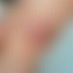
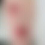
Intolerably severe pruritus. Rapid succession or simultaneous appearance of exudative, reddened, infiltrated, nummular plaques, reminiscent of nummular dermatitis, lichenified papules, or small urticarial papules. The skin lesions are extremely resistant to therapy.
LaboratoryThis section has been translated automatically.
Frequent eosinophilia.
HistologyThis section has been translated automatically.
Perivascular infiltrates from lymphocytes, granulocytes, eosinophils, plasma cells. Swelling of the vascular endothelia (unspecific image).
Differential diagnosisThis section has been translated automatically.
Nummular dermatitis; Scattering contact dermatitis; dermatitis herpetiformis; Lichen planus; Mycosis fungoides; Drug exanthema; Seborrheic dermatitis; Pityriasis rosea;
External therapyThis section has been translated automatically.
Internal therapyThis section has been translated automatically.
Therapy of the 1st choice are glucocorticoids, perorally, in medium dosage, initial 60-80 mg/day prednisolone equivalent, slow release to low maintenance dose over several weeks. After discontinuation the skin symptoms flare up again. The glucocorticoids can only be completely eliminated after months or years.
Cases with healing under exclusive azathioprine therapy (e.g. Imurek) are described. An attempt at therapy with antihistamines such as desloratadine (e.g. Aerius) 1-2 tbl/day or levocetirizine (e.g. Xusal) 1-2 tbl/day is possible.
Progression/forecastThis section has been translated automatically.
Note(s)This section has been translated automatically.
The technical term "-oid" is mainly used to describe a body, a form, a structure or the like that is comparable to something - is similar to something. In this disease, 2 types of efflorescence (e.g. lichenoid and exudative nummular plaques) should always be found that are morphologically similar.
LiteratureThis section has been translated automatically.
- Jansen T et al (1992) Sulzberger's scar exudative discoid and lichenoid chronic dermatosis ("Oid-Oid disease")--reality or fiction? Dermatologist 43: 426-431
- Rongioletti F et al (1989) Exudative discoid and lichenoid chronic dermatosis (Sulzberger's scar) a fictional disease? Int J Dermatol 28:40-43.
- Savill T (1891) On an epidemic skin disease. Br Med J (London) 2: 1197-1202.
- Stevens DM et al (1984) On the concept of distinctive exudative discoid and lichenoid chronic dermatosis (Sulzberger's scar). Am J Dermatopathol 6: 387-395
- Sulzberger MB, Garbe W (1937) Nine cases of a distinctive exudative discoid and lichenoid chronic dermatosis. Arch Dermatol (Chicago) 36: 247-278.
- Tabara K et al (2013) A 6-year-old boy with Sulzberger and Garbe dermatosis: a case report and literature review. Postepy Dermatol Alergol 30:403-8.
Recommended articles


Incoming links (8)
Betamethasone valerate emulsion hydrophilic 0,025/0,05 or 0,1 % (nrf 11.47.); Dermatosis, exudative discoid lichenoid; Dermatosis, exudative discoid lichenoid, chronic; Eosinophilia and skin; Exudative discoid lichenoid dermatitis; Oid-oid-disease; Sulzberger-garbe syndrome; Triamcinolone acetonide cream hydrophilic 0,025/0,05/0,1% (nrf 11.38.);Outgoing links (22)
Adverse drug reactions of the skin; Antihistamines, systemic; Azathioprine; Betamethasone valerate emulsion hydrophilic 0,025/0,05 or 0,1 % (nrf 11.47.); Bloch, bruno; Contact dermatitis (overview); Dermatitis herpetiformis; Desloratadine; Eczema (overview); Glucocorticosteroids systemic; ... Show allDisclaimer
Please ask your physician for a reliable diagnosis. This website is only meant as a reference.