Synonym(s)
HistoryThis section has been translated automatically.
Gibert, 1860; there is evidence that the disease was first described by Robert Willan in 1798 under the name "Roseoal annulata".
DefinitionThis section has been translated automatically.
Frequent, exanthematic, self-limiting, inflammatory skin disease of unknown (probably viral etiology) etiology with typical two-phase course.
You might also be interested in

Occurrence/EpidemiologyThis section has been translated automatically.
Common clinical picture with a prevalence of 0.1%.
EtiopathogenesisThis section has been translated automatically.
Unknown.
The role of the herpes viruses HHV-6(HHV-7), possibly also HHV-8, is increasingly being discussed, especially as their antigens were detectable in the lesions and the disease responds to antiviral substances such as acyclovir.
An association with the H1N1 virus and COVID-19 has been reported several times (Martora F et al. 2022).
Furthermore, the occurrence of a pityriasis rosera-like vaccination reaction after COVID-19 vaccination has been observed (Dormann H et al. 2021; Gambichler T et al. 2022; Cohen OG et al. 2021). In this case, the primary medallion usually formed in the classic variant is absent (Kussini J et al. 2025). Histologically, tissue eosinophilia may be present S.a. under cutaneous vaccination reactions.
Associations with other viruses(EBV, CMV) or streptococcal infections could not be clearly verified.
Other triggers under discussion are:
- Medications (captopril, gold, isotretinoin, non-steroidal anti-inflammatory drugs, omeprazole, terbinafine and tyrosine kinase inhibitors have been associated with the triggering of "pityriasis rosea-like exanthema").
- wearing (unwashed) impregnated clothing
- stress (?).
ManifestationThis section has been translated automatically.
Occurring in healthy individuals between the ages of 10-35 years (Leung AKC et al. 2021). Occurrence of pityriasis rosea in children <10 years of age is uncommon. Females appear to be preferentially affected (m:w = 1:2). Equal occurrence worldwide. Endemic occurrence possible especially in spring and autumn.
LocalizationThis section has been translated automatically.
Trunk (up to the base of the neck) and proximal (approximately upper third) extremities. The head, neck and peripheral extremities remain free. An inverse form(pityriasis rosea inversa) affects the axillae and the groin region.
ClinicThis section has been translated automatically.
Occasionally, mild prodromes such as respiratory symptoms (about 10% of patients), nausea, malaise, lassitude, fever, and headache precede the initial skin lesions. Less commonly, enlarged lymph nodes and arthralgias are detected (in 5% of patients - Leung AKC et al. 2021).
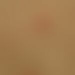
Initially, a 0.4-7.0 cm, oval, light red to pink, usually scaling (Collerette scaling), marginal plaque ("primary medallion") with a somewhat blanched center is seen. This primary medallion (tache mère, herald patch) is a regularly occurring symptom in > 50% of cases, is certainly overlooked many a time, occurs mostly on the trunk, but also on the upper arms or thighs. Mild itching is reported by about 25% of affected individuals. Usually isolated, the primary plaque embodies the only manifestation of this disease in a period of 4-14 days. Thereafter, secondary exanthema occurs in the majority of cases. Multiple primary plaques are possible, but they are rare.
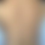
In the further course, relapsing, exanthematous (symmetrical) spreading (truncal), lasting for 1-2 weeks, of 0.2-1.0 cm, oval or elongated, little raised, pink-colored spots with a smooth or finely scaling surface, aligned according to the cleavage lines (fir tree pattern; canopy sign). The secondary efflorescences are also called "medallions". Their number varies, ranging from a few to over a hundred.
Usually they remain isolated, rarely there is confluence of lesions. The chest, neck, and upper arms are affected first, and later the lateral parts of the trunk, abdomen, and proximal upper neck areas. The foci may remain small but may also grow within 7-20 days, then sink into the center and show a dry scaly surface (see Fig.). In individual patches/plaques, evidence of so-called collerette scaling, an inwardly directed delicate scaly ruff, is possible. This type of scaling is suitable as a diagnostic phenomenon (see Fig.). Lichenoid, also vesicular or erythema multiforme-like lesions can be observed (Benedek 1931).
The face, capillitium, and distal halves of the extremities usually remain free of the secondary exanthema (in black populations, involvement of neck areas, face, and capillitium is regularly observed).
Oropharyngeal mucosal lesions are rare but can be attributed to the clinical picture (Ciccarese G et al. 2017). Round, erythematous patches with grayish-white coatings on the anterior and middle buccal mucosa are described; coarser scaling and pinpoint hemorrhages are also possible. Erythematous plaques may also occur on the hard palate.
Rare variants:
- follicular pityriasis rosea
- hemorrhagic and urticarial pityriasis rosea
- circine, vesicular, squamous or psoriasiform pityriasis rosea.
- Inverse pityriasis rosea is observed preferentially in children as well as African-Americans(pityriasis rosea inversa).
- Unilateral pityriasis rosea has been reported (Ghariani Fetoui N et al. (2020).
- Co-infestation of palms and soles has been reported several times (Cabre J et al. 1970)
Pityriasis rosa in colored populations: In dark skin the exanthema appears more infiltrated than in light skin with a clear tendency to hypo but also to hyperpigmentation! Depending on the degree of pigmentation, the characteristic red color of the skin manifestations in white skin is missing. Instead, either rich-brown but also gray-brown shades impress (see Fig.). There were no differences in the time of occurrence and the prevalence of itching or the occurrence of primary daillons. In a larger case series (n=50), the face was affected with 30% and the capillitium with about 8% (this pattern is significantly more frequent than in white populations).
HistologyThis section has been translated automatically.
Nonspecific superficial dermatitis with rather minor acanthosis, irregularly elongated rice ridges, variably intense papilledema, perivascular and interstitial lymphocytic infiltrate; occasional admixture of eosinophilic granulocytes. Marked epidermotropia with focal spongiotic loosening of the epithelium to vesicle formation, focal stratified parakeratosis (histologic counterpart of Collerette's pucker). Rarely, erythrocyte extravasation.
DiagnosisThis section has been translated automatically.
Typical clinical picture with biphasic course, absence of systemic reactions, presence of a primary medulla, biphasic onset, involvement of covered skin areas.
Differential diagnosisThis section has been translated automatically.
- Clinical Differential Diagnosis:
- Tinea corporis: never exanthematic, but chronically continuous course (rare exception is microsphere, here there is self-fluorescence under wood light); pathogen detection by native examination and culture is proof.
- Psoriasis vulgaris: first manifestation of an acute, exanthematic psoriasis as an important DD. The exanthema phenomenon is always negative in pityriasis rosea!
- Parapsoriasis en plaques (benign small-field form): chronic process, never acute exanthematic. Single florescences significantly larger than in Pityriasis rosea. Typical is a "pseudoatrophy of the surface".
- Drug exanthema: Acute monomorphic exanthema; rarely eczematous or psoriasiform. Drug anamnesis with prompt re-prescription of a drug.
- Pityriasis lichenoides: Similar distribution pattern. However, PLEVA and PLC show a groundbreaking polymorphism (colourful picture) of the efflorescences.
- Early syphilis: Syphillis are usually accompanied by swelling of the LK. Distribution pattern: Frequent infestation of palms and face. Serology is conclusive! Histology is conclusive (plasmacellular dermatitis).
- Histological differential diagnoses:
- Acute and subacute eczema: spongiosis, extensive parakeratosis, in atopic eczema possible prominent eosinophilia. Often indistinguishable.
- Psoriasis guttata: acanthosis, extensive hyper- and parakeratosis with neutrophil inclusions, diffuse, also perivascularly compressed lymphocytic infiltrate with neutrophil granulocytes, no erythrocyte extravasations, strong epidermotropism.
- Parapsoriasis en plaques: Fibrosis of the papillary stratum; surface epithelium rather atrophic, epidermotropy present, hardly any spongiosis, no parakeratosis!
- Allergic contact dermatitis: Clinically clearly distinguishable! Prominent, flat spongiosis, surface epithelium acanthotic, long hills of parakeratosis. Histological differentiation can only be reliably made in connection with clinical data.
- Tinea corporis: Varied histological pattern, which is characterized by the acuteity of the infection. In early stages low superficial perivascular lymphocyte infiltrate, focal spongiosis; later stage with neutrophil component. Compact ortho- and parakeratosis. In the PAS preparation, detection of hyphae (then safe DD); otherwise safe DD is only possible in connection with clinical data.
- Erythema anulare centrifugum: Clinically clearly distinguishable; histologically safe differentiation only in connection with clinical data! Mostly dense perivascular infiltrate sheaths.
- Drug exanthema: Cancellous drug exanthema are rare! Mostly combined with interface dermatitis.
- Early syphilis: Interface dermatitis with psoriasiform epidermal reaction dense, band-shaped infiltrate in the upper and middle dermis (lymphocytes, histiocytes and plasma cells. Extension of the infiltrate to the deep vascular plexus).
TherapyThis section has been translated automatically.
General therapyThis section has been translated automatically.
External therapyThis section has been translated automatically.
Cave! Irritation from aggressive external agents. No fat creams or ointments. Treatment e.g. with Tannolact Lotio, Tannosynt Lotio (shaking mixture), Optiderm Lotio. Blande skin care with O/W emulsion (e.g. Eucerin O/W). Recommended especially for itching are creams containing 5% polidocanol or shaking mixture R200 or weakly effective glucocorticoids like 0.5% hydrocortisone emulsion R123.
Radiation therapyThis section has been translated automatically.
Mild broad-spectrum UVB therapy can be applied to fair-skinned patients with extensive infestation (Villalon-Gomez JM 2018).
Internal therapyThis section has been translated automatically.
If necessary, oral antihistamines such as levocetirizine (e.g. Xusal Tbl.) 1 time/day 5 mg p.o. or desloratadine (e.g. Aerius Tbl.) 1 time/day 5 mg p.o.
Alternative: Aciclovir; in a randomized study in a medium-sized collective, a positive clinical effect (shortening of disease duration) was demonstrated by oral administration of aciclovir (OFF label use) for 7 days (400 mg/5xday p.o.). This therapeutic option should be carefully considered with regard to the self-limiting course of the disease.
Alternative: Oral erythromycin (250mg 4x/day for 2 weeks) is significantly successful according to a meta-analysis (Chuh A et al. 2005).
Progression/forecastThis section has been translated automatically.
Spontaneous healing occurs within in 6-12 weeks, usually without persistent skin changes. Hyperpigmentation, leukoderm formation are possible. Sporadic scarring is also possible. Lifelong immunity is assumed. Recurrences are rare (<3%) (pityriasis rosea recidivans).
In a larger study in dark-skinned individuals (n=50), differences in progression and progenosis are evident (Amer A et al. 2007). In nearly 50% of patients, the disease regressed within 4-6 weeks. Persistent hyperpigmentation was observed in about half of the patients, hypopigmentation in about 30%.
Note(s)This section has been translated automatically.
Remember! A primary medallion is a regularly occurring, diagnostically usable symptom; in > 50% of cases
Notice! Pityriasis rosea (almost) never affects the face and mucous membranes; only rarely the extremities! Patients have no disturbed general condition!
Notice! The course of pityriasis rosea is critical for the fetus up to the 15th week of pregnancy with an increased risk (57%) of miscarriage or premature birth (Monastirli A et al. 2016).
LiteratureThis section has been translated automatically.
- Amer A et al. (2007) The natural history of pityriasis rosea in black American children: how correct is the "classic" description? Arch Pediatr Adolesc Med 161:503-506
- Benedek Pityriasis rosea. Arch Derm Syph 24, 557-570 (1931)
- Cabre J et al (1970) Pityriasis rosea Gibert. In: Gottron-Schönfeld, Dermatologie jund Veneroloie, Ergänzungsband S 71-89.Thieme Verlag Stuttgart
- Chuang TY et al. (1983) Recent upper respiratory tract infection and pityriasis rosea: a case-control study of 249 matched pairs. Br J Dermatol 108: 587-591
- Chuh A et al.(2006) Atypical presentations of pityriasis rosea: case presentations. J Eur Acad Dermatol Venereol 19:120-126.
- Ciccarese G et al. (2017) Oropharyngeal lesions in pityriasis rosea. J Am Acad Dermatol 77:833-837.
- Cohen OG et al. (2021) Pityriasis rosea after administration of Pfizer-BioNTech COVID-19 vaccine. Hum Vaccin Immunother 17:4097-4098.
- Das A et al. (2015) Acyclovir in pityriasis rosea: An observer-blind, randomized controlled trial of effectiveness, safety and tolerability. Indian Dermatol Online J 6:181-184
- Demirkan S et al (2019). Does influenza subtype H1N1 have a place in the etiology of pityriasis rosea?. Postepy dermatologii i alergologii 36: 164-166.
- Drago Fet al.(2014) Relapsing pityriasis rosea. Dermatology 229:316-318
- DragoF et al. (2008) Pregnancy outcome in patients with pityriasis rosea. J Am Acad DErmatol 58: 78-83
- Ermertcan AT et al (2010) Childhood Pityriasis rosea inversa without Herald Patch Mimicking Cutaneous Mastocytosis. Iran J Pediatr 20:237-241.
- Gambichler T et al. (2022) Cutaneous findings following COVID-19 vaccination: review of world literature and own experience. J Eur Acad Dermatol Venereol 36:172-180.
- Ghariani Fetoui N et al (2020) Unilateral pityriasis rosea. Int J Dermatol 59:e27-e28.
- Gibert CM (1860) Traité Pratique des Maladies de la peau et de la Syphilis, 3 rd edn. Plon (Paris) pp:402
- Güleç Aİ et al. (2016) Pityriasis rosea-like adverse reaction to atenolol. Hum Exp Toxicol 35:229-231.
- Kempf W (2002) Human herpesvirus 7 in dermatology: what role does it play? Am J Clin Dermatol 3: 309-315
- Kempf W, Burg G (2000) Pityriasis rosea--a virus-induced skin disease? An update. Arch Virol 145: 1509-1520
Kluger N (2023) Annular erythemas and purpuras. Life (Basel) 13:1245.
Kussini J et al. (2025) Cutaneous reactions to vaccination. J Dtsch Dermatol Ges 23:195-209.
- Leung AKC et al. (2021) Pityriasis Rosea: An Updated Review. Curr Pediatr Rev 17:201-211.
- Martora F et al. (2022) Can COVID-19 cause atypical forms of pityriasis rosea refractory to conventional therapies? J Med Virol 94:1292-1293.
- Mayfield J et al.(2020) Childhood Pityriasis Rosea With Multiple Herald Patches. Cureus 12: e9876.
- Monastirli A et al. (2016) Gestational Pityriasis Rosea: Suggestions for Approaching Affected Pregnant Women. Acta Dermatovenerol Croat 24:312-313.
- Polat M et al. (2014) Pityriasis rosea-like drug eruption due to bupropion: a case report. Hum Exp Toxicol 33:1294-129
- Rassai S et al. (2011) Low dose of acyclovir may be an effective treatment against pityriasis rosea: a random investigator-blind clinical trial on 64 patients. JEAV 25: 24-26
- Renner R et al. (2010) Chronic inflammatory and autoimmune-mediated dermatoses in pregnancy. Dermatology 61: 1021-1026
- Vaccaro M et al. (2020) Pityriasis rosea during omalizumab treatment for chronic spontaneous urticaria. Dermatol Ther 33:e14356.
- Vano-Galvan S et al. (2009) Atypical pityriasis rosea in a black child: a case report. Cases J 2:6796.
- Villalon-Gomez JM (2018). Pityriasis Rosea: Diagnosis and Treatment. Am Fam Physician 97:38-44.
- Watanabe T et al. (2002) Pityriasis rosea is associated with systemic active infection with both human herpesvirus-7 and human herpesvirus-6. J Invest Dermatol 119: 793-797
Recommended articles


Incoming links (38)
Annular dermatoses; Asymmetric periflexural exanthema of childhood ; Collerette; Cutaneous vaccination reactions; Dermatitis; Dermatitis exudative discoid lichenoid; Dermatoses, erythematosquamous; Erythema; Erythema anulare centrifugum; Erythrosquamous diseases; ... Show allOutgoing links (33)
Aciclovir; Adverse drug reactions of the skin; Antihistamines, systemic; Atopic dermatitis (overview); Contact dermatitis allergic; Covid-19; Cutaneous vaccination reactions; Cytomegalovirus; Desloratadine; Drug exanthema maculo-papular; ... Show allDisclaimer
Please ask your physician for a reliable diagnosis. This website is only meant as a reference.