Synonym(s)
HistoryThis section has been translated automatically.
DefinitionThis section has been translated automatically.
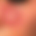
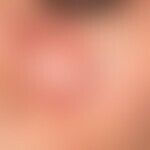
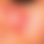
Most frequent form of cutaneous lupus erythematosus with circular, oval or garland-shaped, red plaques with flat, lesional, mostly small-spotted adherent scaling (which may also be absent). Characteristic follicular hyperkeratosis can occur when scaling is present. Typical, and thus diagnostically groundbreaking for CDLE, is scarring, which usually occurs in the centre of the lesion. In older lesions the circumscribed scarring may dominate the clinical picture. In older lesions, the scarring can dominate the clinical picture, and red or whitish, scaly plaques can cover a light scar area. The healed lesion of chronic discoid lupus erythematosus is a non-irritant, sunken scar. The process of scarring healing can take years.
Hypertrophic chronic discoid lupus erythematosus ( lupus erythematosus verrucosus) is to be regarded as a variant.
You might also be interested in

Occurrence/EpidemiologyThis section has been translated automatically.
Incidence: 15-50/100.000 inhabitants/year. Worldwide occurrence. African Americans have a higher prevalence than Caucasian Americans. Asians show similar prevalence to Caucasians.
EtiopathogenesisThis section has been translated automatically.
Unknown. Autoimmune disease with genetic predisposition. Provocation and entertainment of manifestation by exogenous factors such as trauma, light, cold, stress, infections.
ManifestationThis section has been translated automatically.
Mainly occurring in adults (20 to 40 years of age).
f:m=3:1
Rarely heredity exists.
LocalizationThis section has been translated automatically.
Mostly face (cheeks, forehead, nose), auricles, but also hairy head, chest, rarely trunk and extremities.
ClinicThis section has been translated automatically.
Sharply demarcated, 1.0-10.0 cm large, solitary, few or multiple, homogeneously reddened, scaly, usually only slightly elevated, but clearly indurated red plaques. Peripheral growth may result in circular, sometimes annular, structures with a flaking scarred center(atrophy of follicular ostia; look for it carefully!) and peripheral zone of progression. Possibly confluence of the foci. Typical is a hypersensitivity of the inflammatory zone (recommendation: brush with light spatula pressure from the healthy skin into the diseased skin). In the scarring center of the discoid foci, depigmentation, hyperpigmentation and/or telangiectasia are also seen. Solitary or multiple, hypersensitive foci of alopecia with central scarring and, depending on the activity, peripheral inflammatory progression zone appear at the capillitium. S.a.u:
- Alopecia, scarring
- Lupus erythematosus tumidus
- Chilblain's lupus
- Lupus erythematosus profundus
- Lupus erythematosus verrucosus.
Characteristic of discoid lupus erythematosus on reflected light microscopy are follicular hyperkeratosis in the foci, and hyperesthesia when the foci are touched(tapestry nail phenomenon).
LaboratoryThis section has been translated automatically.
Mostly without pathological findings. Rarely leukopenia and positive (low-titer) antinuclear antibodies (ANA). It is important to exclude SLE (see below lupus erythematosus, systemic).
HistologyThis section has been translated automatically.
Direct ImmunofluorescenceThis section has been translated automatically.
In affected skin detection of IgG, IgM, IgA,C1 andC3 at the dermo-epidermal junction zone (= positive lupus band test); unaffected skin is negative.
Differential diagnosisThis section has been translated automatically.
External therapyThis section has been translated automatically.
Exclusively external measures are not always sufficient!
- Consistent light protection. Use of sunscreens with a high protection factor, UVA and B filters (preferably sun protection preparations on a physical basis) and consistent textile sun protection. It is important to inform the patient comprehensively about the various sun protection measures.
- Topical glucocorticoids. Depending on the localisation and activity of the HV, an appropriate class of active substances is selected. Possibly occlusive dressings.
- Capillitium: Steroid-containing solution. (e.g. Ecural-Lsg.) once/day, if necessary additional injection with glucocorticoid crystal suspension( triamcinolone acetonide, e.g. Volon A, 10-20 mg together with 2 ml 1% mepivacaine and applied several times with a thin cannula at intervals of 4-6 weeks intralesionally).
- Topical immunomodulators: In pilot studies good effects by local treatment with tacrolimus (e.g. 1% protopic ointment) or Pimecrolimus (e.g. Elidel).
Internal therapyThis section has been translated automatically.
Antimalarials: Hydroxychloroquine (Quensyl®) or chloroquine (Resochin®) are the first choice! They show satisfactory results in 50-90% of patients. Dosage of chloroquine: initially 4.0 mg/kg bw/day; children: 3.5 mg/kg bw/day. After one month disease-adapted reduction of the dose. Regular ophthalmologic check-ups due to possible retinopathy. Chloroquine leads to a reduction of HLA-DR+/CD1a+ cells in lesional skin.
Alternative: hydroxychloroquine (Quensyl®, dosage: 6.5 mg/kg bw/day; children: 6.0 mg/kg bw/day), disease-adapted reduction of the dose. Total dose 100 mg. In individual cases with good tolerance, the total dose can be significantly exceeded.
Please note! Smokers respond significantly less well to treatment with antimalarials than non-smokers! Pregnancy with antimalarials is not a contraindication. Note: Antimalarials such as cloroquine and hydroxychlorquine can trigger hemolytic crises in the presence of glucose-6-phosphate dehydrogenase deficiency. In this respect, the patient should be asked about this (FA; signs of favism).
If necessary, combine with glucocorticoids (e.g. prednisolone 5 mg/day p.o.).
In case of extensive findings and lack of response to other therapies glucocorticoid shock therapy: Initially 100 mg/day prednisolone (e.g. Solu-Decortin H) i.v.; slowly taper off. Maintenance dose: Prednisolone 10-20 mg/day (e.g. Decortin® H) p.o. If necessary, combination with azathioprine (e.g. Imurek ®50-100mg/day) p.o.
Retinoids such as acitretin (neotigason 10-20 mg/day) are used as an alternative to chloroquine. The therapeutic successes are not particularly convincing.
Studies with methotrexate (intravenous therapy at 15-25 mg/week) and mycophenolate mofetil (MMF) show good clinical success. MMF is used in particular in patients who are refractory to antimalarials. MMF can be used alone or in combination with antimalarials in a dosage of between 1500 and 3500 mg p.o./day.
Thalidomide: can be used as an alternative in severe cases of CDLE (Chasset et al. 2018). A high response rate (90%) is offset by high rates of treatment discontinuation (24%) due to side effects (neuropathy, thromboembolic processes).
Operative therapieThis section has been translated automatically.
- Physical or surgical measures should be considered after weighing the risk-benefit ratio against other procedures. To be considered:
- Cryosurgery in open or closed procedures; for stamping procedures, cool the stamp surface to 0 °C, place the stamp loosely and freeze briefly until the stamp temperature (not tissue temperature) of -150 °C is reached. Repeat the procedure immediately after thawing.
- Cauterization: Lesions can also be carefully coagulated with electrocautery; surface scabbed, post-cure with sharp curette (boot curette no. 6).
Progression/forecastThis section has been translated automatically.
Chronic course, acute exacerbation with visceral involvement possible; quod vitam favourable, defect healing. A transition to systemic lupus erythematosus is reported in about 1% (other sources say 5-10%!!) of cases.
Note(s)This section has been translated automatically.
Many patients report light sensitivity, possibly burning and itching after UV exposure.
However, a smaller number of patients seem to benefit from sun exposure.
LiteratureThis section has been translated automatically.
- Bohm I et al (1998) ANCA-positive lupus erythematodes profundus. Successful therapy with low dosage dapsone. dermatologist 49: 403-407
- Calza L et al (2003) Systemic and discoid lupus erythematosus in HIV-infected patients treated with highly active antiretroviral therapy. Int J STD AIDS 14: 356-359
- Cazenave PL (1844) Pemphigus chronique, générale; forme rare de pemphigus foliacé; mort: autopsy; alteration du foie. Les Annales des maladies de la peau et de la syphilis 18: 583-585
- Chasset F et al (2018) Efficacy and tolerance profile of thalidomide in cutaneous lupus erythematosus:
Asystematic review and meta-analysis. J Am Acad Dermatol 78:342-350 - Costedoat-Chalumeau N et al (2005) Safety of hydroxychloroquine in pregnant patients with connective tissue disease. Autoimmune Rev 4: 111-115
- Gammon B et al (2011) Efficacy of mycophenolate mofetil in antimalarial-resistant cutaneous lupus erythematosus. J Am Acad Dermatol 65:717-21
- Günther C (2015) Genetics of lupus erythematosus. dermatologist 66: 121-130
- Jewell ML et al (2000) Patients with cutaneous lupus erythematosus who smoke are less responsive to antomalaria treatment. J Am Acad Dermatol 42: 983-987
- Kreuter A et al (2004) Pimecrolimus 1% cream for cutaneous lupus erythematosus. J Am Acad Dermatol 51: 407-410
- Marzano AV et al (2014) Drug-induced lupus erythematosus. G Ital Dermatol Venereol 149:301-309
- Moises-Alfaro C et al (2003) Discoid lupus erythematosus in children: clinical, histopathologic, and follow-up features in 27 cases. Pediatric dermatol 20: 103-107
Parodi A et al (2014) Cutaneous manifestations of lupus erythematosus. G Ital Dermatol Venereol 149:549-554
- Wenzel J et al (2005) Efficacy and safety of methotrexate in recalcitrant cutaneous lupus erythematosus: results of a retrospective study in 43 patients. Br J Dermatol 153: 157-162
- Wimmershoff MB et al (2003) Discoid lupus erythematosus and lupus profundus in childhood: a report of two cases. Pediatric dermatol 20: 140-145
Recommended articles


Incoming links (32)
Atrophoderma vermiculatum; Bowen's disease; Camouflage; Cdle; Chloroquine; Complement component 5 deficiency; Dermatoses, erythematosquamous; Discoid lupus erythematosus; Dle; Frontal fibrosing alopecia; ... Show allOutgoing links (40)
Acitretin; Actinic keratosis; Alopecia scarring; Antimalarials; Apoptosis; Autoimmune diseases; Cauterization; Chilblain lupus; Cryosurgery; Depigmentation; ... Show allDisclaimer
Please ask your physician for a reliable diagnosis. This website is only meant as a reference.


 blephari...")
 blephari...")