Synonym(s)
DefinitionThis section has been translated automatically.
Benign, circumscribed, nodular neoplasms of the subcutaneous fatty tissue, occurring singly or multiply. Lipomatoses are rare, syndromal, large-volume fatty tissue hamartomas.
ClassificationThis section has been translated automatically.
Depending on their localization in the skin and in deeper tissues as well as on various other clinical aspects, they can be subdivided:
- Solitary or multiple non-syndromal lipomas:
- Subcutaneous (in loco typico) lipoma
- Special form: Lipomatosis dolorosa
- Dermal lipoma (rarity) and its "hamartoma variant" = Naevus lipomatodes cutaneus superficialis (Hoffmann-Zurhelle)
- Subfascial lipoma (forehead-hairline)
- Intramuscular lipoma (thigh/trunk)
- Spindle cell lipoma/pleomorphic lipoma
- synovial lipoma
- Hibernoma
- Lipoblastoma
- Subcutaneous (in loco typico) lipoma
- Solitary or multiple syndromal lipomas:
- Lipomatoses (see below lipomatosis benign symmetrical)
- Lumbosacral lipoma (often associated with spina bifida)
- Gorlin Goltz Syndrome
- Pringle-Bourneville phacomatosis
- Wermer Syndrome
- Gardner Syndrome
- Proteus Syndrome
You might also be interested in
EtiopathogenesisThis section has been translated automatically.
ManifestationThis section has been translated automatically.
LocalizationThis section has been translated automatically.
The classic lipoma is mainly found on the trunk, here mainly on the shoulders, arms and thighs.
Smaller lipomas in the forehead area (frontalis-associated lipomas), which are cosmetically disturbing, are rarer.
Clinical featuresThis section has been translated automatically.
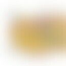
Solitary or multiple (2-3% of cases), visible or only palpable, soft or bulging, round or elongated, easily delimitable, 2.0-5.0 cm in diameter (exceptionally up to 10 cm), mostly subcutaneously located, soft or firm tumour, which can be displaced and delimited from deeper structures. The lipoma often bulges the overlying skin hemispherically and thus visibly forms contours. Subjectively, lipomas are mostly asymptomatic. Occasionally pain (due to pressure on nerve endings?).
Special forms:
- Multiple painful lipomas are called lipomatosis dolorosa (mainly obese women during and after menopause).
- The subfascial lipoma is found as a solitary, harmless skin-coloured, firm nodule (or knot) on the forehead (forehead lipoma).
- Intramuscular lipomas occur in adults mainly in the shoulder and pelvic area, as painless soft swelling of the tissue.
Isolated malformations of the adipose tissue:
- The mostly large-volume fatty tissue hamartomas are called lipomatoses and occur as a partial symptom of benign symmetrical lipomatosis.
- The rare fatty tissue hamartoma localized in the dermis is called nevus lipomatosus cutaneus superficialis.
HistologyThis section has been translated automatically.
- Histological variants:
- Angiolipoma (see also angiolipomatosis) and its variant the cellular angiolipoma
- Spindle zellipoma
- Pleomorphic lipoma
- Sclerotic lipoma
- Myolipoma (admixture of smooth musculature; especially in the pelvic area in women)
- Angiomyolipoma (more common in the kidney in Pringle-Bourneville phacomatosis; see also angiolipomatosis, familial)
- Angiomyxolipoma
- Chondroid lipoma.
- As morphological and clinical peculiarity the hibernoma and the lipoblastoma occurring in childhood are to be addressed. The pleomorphic lipoma is largely identical to the spindle cell lipoma and shows as histological feature flower-like giant cells (giant cells with an eosinophilic centre).
- In the sclerotic lipoma, a connective tissue parenchyma, in places with a storiform pattern, into which ripe adipocytes are scattered (picture of a Swiss cheese).
- In the case of myolipoma there are different admixtures of smooth musculature.
- The angiomyolipoma, predominantly occurring as a renal tumor in the context of Pringle-Bourneville phacomatosis, more rarely in other organs (including the skin), shows, in addition to a vascular component, a varying proportion of smooth musculature, partly with epithelioid character (positive for HMB45!).
- In the chondroid lipoma, chondroid differentiations are found in the tumor parenchyma. The mature adipocytes indicate the benignity of the tumor.
Differential diagnosisThis section has been translated automatically.
TherapyThis section has been translated automatically.
In case of cosmetic or mechanical disorder, or in case of permanent pain, extirpation.
Alternatively, liposuction is also possible for larger lipomas, the results require further scientific evaluation.
Alternative: Deoxycholic acid/polydenylphosphatidcholine. Injection treatments with phospholipids (Lipostabil®) are still considered to be experimental. They should only be performed by physicians who are familiar with this procedure. The injections are performed with a needle size between 21 G and 25 G. The injection is carried out quantitatively until a resistance becomes noticeable (5.0 ml injection solution for small to medium-sized lipomas) . Treatment is carried out every 8-12 weeks. Usually 2-3 treatments are necessary.
LiteratureThis section has been translated automatically.
- Aydogdu E et al (2004) Giant lipoma of the back. Dermatol Surgery 30: 121-122
- Bardazzi F et al (2003) Subungual lipoma. Br J Dermatol 149: 418
- Barde NG et al (2014) Frontalis-associated lipoma: a rare casereport
. Indian J Dermatol 59:96-98 - Brass D et al (2015) Successful treatment of forehead lipoma depends on knowledge of the surgical anatomy: a step-by-step guide. Clin Exp Dermatol. doi: 10.1111/ced.12741
- Choi HS et al (2003) Lipoma of the index finger. J Dermatol 30: 834-836
- Mentzel T (2000) Lipomatous tumors of the skin and soft tissue. New entities and concepts. Pathologist 21: 441-448
- Wilk M et al (2018) Mesenchymal and neuronal tumors. In: Plewig G et al (ed.) Braun-Falco`s Dermatology, Venerology and Allergology. Springer publishing house SS 1887-1919
- Zelger B (2003) Tumours of the nerves, muscles, cartilage, bones and fatty tissue of the skin. In: Kerl H et al (eds.) Histopathology of the skin. Springer Verlag, Berlin Heidelberg New York, S. 848-855
Recommended articles


Incoming links (34)
Adiposis dolorosa; Angiolipomatosis, familial; Angiomyolipoma; Angiomyxoma cutaneous; Bannayan-riley-ruvalcaba syndrome; Biker's nodules; Birt-hogg-dubé syndrome; Cloves syndrome; Cowden syndrome; Elastofibroma; ... Show allOutgoing links (18)
Adiposis dolorosa; Angiolipoma; Angiolipomatosis, familial; Angiomyxolipoma; Basal cell nevus syndrome; Cyst; Fibroma; Gardner syndrome; Hibernoma; Lipomatosis benign symmetric (overview); ... Show allDisclaimer
Please ask your physician for a reliable diagnosis. This website is only meant as a reference.
Authors
Last updated on: 29.10.2020
Authors: ![]() Prof. Dr. med. Peter Altmeyer Philipp Stüven
Prof. Dr. med. Peter Altmeyer Philipp Stüven
Editor: ![]() Julian Baur
Julian Baur