Synonym(s)
HistoryThis section has been translated automatically.
DefinitionThis section has been translated automatically.
Rare, solitary, rapidly growing, intermediate-malignant, fibrohistiocytic tumor of the skin within a few months with a relatively benign clinical course, which occurs almost exclusively in light-damaged skin of older people. It is probably not an independent clinical picture, but a reaction pattern of different, largely dedifferentiated neoplasias, but also degenerative processes. This fine tissue reaction pattern conceals squamous cell carcinomas, melanomas, basal cell carcinomas, pseudosarcomatous xanthogranuloma variants, etc. Some authors consider atypical fibroxanthoma to be a cutaneous variant of malignant fibrous histiocytoma (Cooper JZ et al. 2005).
You might also be interested in

Occurrence/EpidemiologyThis section has been translated automatically.
The incidence is 2.5/100,000; m>w;
EtiopathogenesisThis section has been translated automatically.
UV as well as previous radiation therapy with X-rays appear to play a decisive pathogenetic role; furthermore, persistent immunodeficiency (increase in AFX in organ transplant patients) as well as genetic defects such as those in xeroderma pigmentosum.
Mutations in the tumor suppressor gene p53, which are thought to be pathogenetically relevant, have been increasingly found. Furthermore, telomerase reverse transcriptase (TERT) promoter mutations have been detected. These mutations lead to activation of telomerases and immortalization of the cells (Grievank KG et al. 2014).
Histogenesis is still unclear: it is likely that the disease originates from poorly differentiated mesenchymal cells of origin, possibly histiocytes or fibroblasts. Proliferation of dedifferentiated keratinocytes (focal keratin expression has been detected in individual cases), myoblastoid proliferation, proliferation of Langerhans cells, etc. are also suspected.
Long-term use of hydrochlorothiazide is thought to be an increased risk factor for atypical fibroxanthoma and pleomorphic dermal sarcoma (Kuntz T et al. 2024).
ManifestationThis section has been translated automatically.
The initial manifestation is particularly frequent during two age peaks, in the 4th and 7th decade respectively, with the latter being significantly larger (in a larger study - Mahalingam S. et al.- the average age was 75.9 years). The first peak refers mainly to non-light-damaged areas (extremities, trunk).
LocalizationThis section has been translated automatically.
Especially ears, nose, cheeks, neck, capillitium. In younger people also extremities or trunk.
ClinicThis section has been translated automatically.
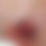
In actinically pre-damaged skin, there is a solitary, up to 3.0 cm large, coarse, indolent, skin-colored, red or brown-red, firm knot with a smooth atrophic surface, which often cannot be displaced on its base (infiltration of the subcutaneous fatty tissue). Often ulcerated and thus encrusted or diffusely bleeding surface.
HistologyThis section has been translated automatically.
Usually well-demarcated nodule confined to the dermis and not extending into the subcutaneous fatty tissue (in contrast to pleomorphic dermal sarcoma), consisting of atypical, spindle-shaped or pleomorphic cells with vesicular or bizarre, hyperchromatic nuclei. Bizarrely configured multinucleated giant cells and atypical mitoses are frequently found.
A clear immunohistologic marker for a definitive diagnosis of atypical fibroxanthoma is still lacking. The cells are CD34, S100, desmin, HMB-45 (human melanoma black 45) negative (Koch M et al. 2015) (DD: dermatofibrosarcoma protuberans) and constantly vimentin positive. Furthermore, there is an inconstant reactivity for alpha smooth muscle actin and for CD68. Occasionally, focal keratin expression is observed (so-called keratin-positive AFX). Furthermore, numerous vascular incisions are found. Frequent focal hemorrhages. Cellular variants with pigmentation and granular or clear cell degeneration are possible.
Myxoid sections, which can also dominate the histological picture, are not uncommon. Molecular pathology: the detection of p53 mutations and TERT promoter mutations provide evidence of UV induction of the tumor.
DiagnosisThis section has been translated automatically.
The clinical findings are not very specific. The histological pattern and especially immunohistochemistry is diagnostically important for AFX.
Differential diagnosisThis section has been translated automatically.
- Clinical:
- Basal cell carcinoma: Like AFX, also in light-exposed areas; typical pearl-like border accentuation of the BCC is missing.
- Spinocellular carcinoma: Clinically indistinguishable! Histology is diagnostic.
- Hyperplastic actinic keratosis: only minimally elevated, clearly increased in consistency by horn deposits. Mostly multitudinous.
- Keratoakanthoma: Keratoakanthoma-like AFX are described. Growth however slow; no central horn crater!
- Granuloma teleangiectaticum: Significantly faster growth; typically not preferred in light exposed areas.
- Malignant melanoma: AFX is unpigmented.
- Merkel cell carcinoma: Fast growing tumour, clinically difficult to assess. Histology is diagnostic.
- Dermatofibrosarcoma protuberans: Age peak of DFSP between the ages of 20 and 40 years. No preference for light-exposed skin areas. Rough, almost woody consistency.
- Skin metastases: Rapid growth is generally expected here; no preference for light-exposed skin areas.
- Histological:
- Carcinoma, spinocellular, desmoplastic: Fine strands or smaller tumor cell nests with atypical, very polymorphic keratinocytes surrounded by a broad desmoplastic stroma. The tumour tends to infiltrate along autochthonous structures such as nerves or vessels. Tumor cells are cytokeratin - and occasionally vimentin - positive.
- Melanoma, malignant, desmoplastic: Fascicular or nodular cell trains of atypical, hyperchromatic, spindle cells located between fibrotically dense collagenous fibres. Occasional mitoses. Desmoplastic melanomas show neurotrophy and follow the course of the dermal nerves (desmoplastic-neutral melanoma). Immunohistology: S-100-, (HMB-45-, Melan A- are not always positive) and vimentin positive (!); S-100 is the most reliable marker.
- Undifferentiated pleomorphic sarcoma (UPS): The AFX is probably the superficial variant of the UPS.
- Malignant fibrous histiocytoma (MFH): This "controversial" tumor species should not be a true differential diagnosis, because UPS and MFH are probably identical tumors (see note).
- Leiomyosarcoma: reactivity for smooth muscle actin and (inconstant) desmin. h- caldesmon +; cytokeratin +/-.
Complication(s)(associated diseasesThis section has been translated automatically.
Tendency to local recurrence (7-9%). These usually occur within a period of 1-2 years. Very rarely metastasis (0.5-4%) mostly in the regional lymph nodes followed by liver, lung and subcutis.
TherapyThis section has been translated automatically.
Excision with a safety margin of at least 0.5 cm. Microscopically controlled surgery is now considered "state of the art".
Radiation therapyThis section has been translated automatically.
Radiation therapy can be used as an alternative in cases of unfavourable surgical site. Response only at higher doses (about 60-65 Gy).
Progression/forecastThis section has been translated automatically.
Favourable prognosis with complete excision on all sides. A metastasis is rare (note: older statistics show a metastasis rate between 0,5%-10%).
AftercareThis section has been translated automatically.
Follow-up should be every six months for the first two years. In the third to the fifth year, annually. Follow-up includes clinical examinations of the patient.
Note(s)This section has been translated automatically.
LiteratureThis section has been translated automatically.
- Cooper JZ et al. (2005) Metastasizing atypical fibroxanthoma (cutaneous malignant histiocytoma): report of five cases. Dermatol Surg 31: 221-255
- Eckert F et al. (1990) The atypical fibroxanthoma. Dermatol 41: 39-42
- Fretzin DF, Hellwig EB (1973) Atypical fibroxanthoma of the skin. Cancer 31: 1541
- Griewank KG et al. (2014) TERT promoter mutations are frequent in atypical fibroxanthomas and pleomorphic dermal sarcomas. Mod Pathol 27:502-508.
- Helwig EB (1963) Atypical fibroxanthoma. Tex J Med 59: 664-667
- Hilgers M et al (2014) Atypical fibroxanthoma on the capillitium. Dermatologist 65: 1008-1010
- Hügel H (2006) Fibrohistiocytic skin tumors. J Dtsch Dermatol Ges 4: 544-555
- Koch M et al. (2015) Atypical Fibroxanthoma - Histological Diagnosis, Immunohistochemical Markers and Concepts of Therapy. Anticancer Res 35:5717-5735.
- Kuntz T et al. (2024) Hydrochlorothiazide and increased risk of atypical fibroxanthoma and pleomorphic dermal sarcoma. J Dtsch Dermatol Ges 22:513-519.
- Mahalingam S et al. (2015) Atypical Fibroxanthoma: A case series and review of literature. Auris Nasus Larynx. 42:469-471
- Rizzardi C et al. (2003) Atypical fibroxanthoma and malignant fibrous histiocytoma of the skin. Anticancer Res 23: 1847-1851
- Stepanova A et al. (2005) A rare low-grade malignant scalp tumor Atypical fibroxanthoma. Dermatology 56: 679-683
Recommended articles


Incoming links (13)
Atypical fibroxanthoma; Cd10; Cutaneous sarcomas (overview); Desmin; Fibrosarcoma paradoxes; Histiocytoma malignant fibrous; Pleomorphic dermal sarcoma; Pleomorphic dermal sarcoma; Pseudosarcoma of the skin; Spindle cell squamous cell carcinoma; ... Show allOutgoing links (26)
Actinic keratosis; Basal cell carcinoma (overview); Carcinoma spinocellular desmoplastic; CD classification; Cytokeratins; Dermatofibrosarcoma protuberans (overview); Desmin; Excision; Histiocytoma malignant fibrous; Hydrochlorothiazide; ... Show allDisclaimer
Please ask your physician for a reliable diagnosis. This website is only meant as a reference.