Synonym(s)
HistoryThis section has been translated automatically.
Laugier and Woringer 1963; Mehregan et al. 1967.
DefinitionThis section has been translated automatically.
Rare chronic skin disease characterized by transepidermal elimination of collagen and/or elastin through the skin. In the acquired (acquired collagenosis) form occurring in adults, it is often associated with diabetes mellitus and/or chronic terminal renal insufficiency.
Basically, there are 2 clinical syndromes:
- The much more common acquired reactive collagenosisin adults, which occurs mainly in diabetics and patients with renal insufficiency (incidences of up to 11% have been described). Acquired reactive collagenosis has also been described in patients with acute myeloid leukemia and as an ADR after treatment with biologics (e.g. tyrosine kinase inhibitors/Soto-García D et al. 2024).
- The very rare familial (primarily described as reactive perforating collagenosis), focal connective tissue degeneration with transepithelial shedding of collagen fibers, which already occurs in children. It can be assumed that this genodermatosis is identical to the"hyperkeratosis follicularis et parafollicularis in cutem penetrans" described by Kyrle.
You might also be interested in

EtiopathogenesisThis section has been translated automatically.
1) Genodermatosis with unexplained aetiopathogenesis.
2) Acquired with underlying diseases such as chronic terminal renal insufficiency or diabetes mellitus. In individual cases, the association with malignancies has been described - prostate carcinoma, chronic lymphocytic leukemia - (Huseynova L et al. 2020)
3) Drug-induced (e.g. inhibitors of the epidermal growth factor EGF/gefitinib, cetuximab/ Vega Díez D et al. 2020), tyrosine kinase inhibitors(nilotinib), monoclonal antibodies against the VEGF receptor(bevacizumab). The multikinase inhibitors sorafenib and cabozantinib have also been shown to cause APD (Soto-García D et al. 2024). Note: Cabozantinib inhibits both the VEGF and EGF signaling pathways. Both are important for vascular homeostasis (Chiang B et al. 2020). EGF and VEGF signals interact additively in the RAS-RAF and PI3K cascades (Lederhandler M et al. 2018).
4) Infections: The clinical picture has also been described in scabies and zoster, whereby the triggering of the disease in these cases is more likely to be reactive as a "Köbner phenomenon" with a corresponding disposition or as an isotopic reaction.
Remark: probably as a result of mechanical or inflammatory irritation of receptive skin, focal damage to the collagenous connective tissue of the skin occurs (scratching can experimentally trigger RPK - Köbner phenomenon), which is finally discharged transepidermally.
Proteolytic enzymes such as matrix metalloproteases or serine proteases as well as modifications of sugar side chains on extracellular matrix proteins such as collagen I and III play an important role.
ManifestationThis section has been translated automatically.
In classic reactive perforating collagenosis, the mean age of onset is 50-60 years. The average duration of disease is 8 months.
LocalizationThis section has been translated automatically.
Mainly extensor sides of the extremities and the trunk. The face, palms of hands and soles of feet remain free of symptoms, as do the mucous membranes.
ClinicThis section has been translated automatically.
There is moderate to marked (non-histaminergic) itching.
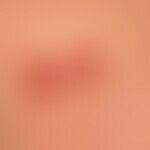
Clinically, there are usually symmetrically distributed, few to numerous, disseminated or linearly grouped ( Köbner phenomenon), initially about 0.2 cm in size, subsequently slowly growing to 1.0 -2.0 cm in size, red, firm papules or plaques with central ulceration and a hard, firmly adherent keratotic plug.
Typical is the detection of a central crusty defect already in initial papules. After its detachment, an ulcerated crater is found, which heals spontaneously after 2-4 weeks with hyperpigmentation.
Note: Although a rare disease, the clinical picture is unique and therefore unmistakable, so that the diagnosis is relatively easy to make if the clinical picture is known
HistologyThis section has been translated automatically.
Hyperplastic epidermis with central bowl-shaped exulceration. In the material stored here, there are horny masses, granulation tissue with triggered, basophilic, collagenous fiber bundles (TOE stain). In the underlying dermis, a nonspecific mixed inflammatory infiltrate is seen.
Differential diagnosisThis section has been translated automatically.
Prurigo simplex subacuta (most important differential diagnosis); no causative underlying disease. Eminently severe itching of the scratched papules.
Prurigo nodularis (see chronic prurigo below)
Rare differential diagnoses:
- Elastosis perforans serpiginosa (anular grouped papules with adherent keratoses)
- Perforating granuloma anulare (skin-colored forked papules)
- Calcinosis cutis (rock-hard, pointed calcium deposits)
- Hyperkeratosis follicularis et parafollicularis in cutem penetrans (mainly localized on the legs)
- Ecthyma (mainly lower leg)
Lues maligna
TherapyThis section has been translated automatically.
A causal therapy is otherwise not known. In the acquired forms, the treatment of the underlying disease is in the foreground (especially diabetes control!).
Externa: Highly potent corticoid externa or external retinoid (the sympt. Therapy with highly potent corticosteroids (also over large areas) has proved successful in our own experience and in the experience of some authors. Injections with depot steroids every 3 weeks are a good alternative, especially in areas of the back that are difficult to reach with cream.
Try PUVA therapy.
Alternative: Good experiences have been made with systemically administered allopurinol (100 mg/day/p.o.). The mechanism of action is unclear. It is possible that the inhibition of xanthine oxidase and an antioxidant effect inhibit the cross-linking of collagen fibers.
Alternative: Recently, several publications have been published describing good clinical results with dupilumab (Gil-Lianes J et al. 2022; Ying Y et al. 2022 )
Care: in addition to consistent skin care, it is advisable to cover the areas with a hydrocolloid plaster (dissolves the central plug, promotes healing, prevents scratching).
Progression/forecastThis section has been translated automatically.
The acquired form of perforating collagenosis is known to have a relapsing course. There is a not inconsiderable tendency to heal spontaneously. It is important to treat the underlying disease (e.g. precise control of diabetes mellitus).
Note(s)This section has been translated automatically.
Perforations of the skin (perforating dermatoses) are found as a secondary phenomenon in a larger number of diseases.
For example: calcinosis cutis, foreign body granulomas, gout, chondrodermatitis nodularis chronica helicis, granuloma anulare, sarcoidosis, necrobiosis lipoidica, malignant melanoma, T-cell lymphoma, Paget's disease, Kyrle 's disease and others.
LiteratureThis section has been translated automatically.
- Chiang B et al. (2020) Rare case of acquired perforating dermatosis induced by cetuximab. J Dermatol 47:e11-e12.
- Fretzin DF, Beal DW, Jao W (1980) Light and ultrastructural study of reactive perforating collagenosis. Arch Dermatol 116: 1054
- Gil-Lianes J et al. (2022) Reactive perforating collagenosis successfully treated with dupilumab. Australas J Dermatol 63: 398-400.
- Huseynova L et al. (2020) Acquired reactive perforating collagenosis in association with prostate adenocarcinoma, chronic lymphocytic leukemia, and Graves' disease. An Bras Dermatol 95:336-339.
- Karpouzis A et al. (2010) Acquired reactive perforating collagenosis: current status. J Dermatol 37:585-592
- Karpouzis A et al. (2004) Acquired reactive perforating collagenosis associated with myelodysplastic syndrome evolving to acute myelogenous leukaemia. Australas J Dermatol 45:78-79
- Kruger K et al (1999) Acquired reactive perforating dermatosis. Successful treatment with allopurinol in 2 cases. Dermatologist 50: 115-120
- Lederhandler M et al. (2018) Unusual eruption in association with sorafenib: a case of acquired perforating dermatosis, reactive perforating collagenosis type. Dermatol Online J 24:13030/qt8b40k5kg.
- Lotz C et al. (2014) Successful treatment of acquired reactive perforating dermatosis with allopurinol. Act Dermatol 4: 84-87
- Mehregan AM, Schwartz OD, Livingood CS (1967) Reactive perforating collagenosis. Arch Dermatol 96: 277
- Rapini RP et al.(1989) Acquired perforating dermatosis. Arch Dermatol 125: 1074 - 1078
- Soto-García D et al. (2024) Acquired perforating dermatoses and cabozantinib: A novel cutaneous toxicity. J Dtsch Dermatol Ges 22:594-596.
- Theodoridis A et al. (2013) Malignant atrophic papulosis (Köhlmeier-Degos disease) - a review. Orphanet J Rare Dis 8:10.
- Vega Díez D et al. (2020) Reactive perforating collagenosis: a rare side effect associated with sorafenib. Rev Esp Enferm Dig 112:960-961.
- Weiss SC (2003) Cutaneous mucormycosis secondary to acquired reactive perforating collagenosis. Cutis 72: 119-123
- Ying Y et al. (2022) Dupilumab may be an alternative option in the treatment of acquired reactive perforating collagenosis combined with AD. Immune Inflamm Dis 10: e574.
Recommended articles


Incoming links (13)
Collagenoma perforans verruciformis; Collagenosis, familial, reactive, perforating; Dermatosis perforating; Elastosis perforans serpiginosa; Folliculitis perforating; Granuloma anulare perforans; Koebner phenomenon; Kyrle's disease; Prurigo simplex subacuta; Reactive perforating collagenosis; ... Show allOutgoing links (26)
Allopurinol; Bevacizumab; Cabocantinib; Calcinosis cutis (overview); Cetuximab; Chondrodermatitis nodularis chronica helicis; Chronic prurigo; Cutaneous t-cell lymphomas; Ekthyma; Elastosis perforans serpiginosa; ... Show allDisclaimer
Please ask your physician for a reliable diagnosis. This website is only meant as a reference.