Synonym(s)
HistoryThis section has been translated automatically.
Winkler 1915
DefinitionThis section has been translated automatically.
Chronic, benign nodule on the free edge of the auricle, which is very painful under pressure. The clinical picture usually persists for 10-15 months before the medical consultation.
You might also be interested in

Occurrence/EpidemiologyThis section has been translated automatically.
Prevalence data are not available. Men are mainly affected (>60%).
EtiopathogenesisThis section has been translated automatically.
Unknown. Probably pressure necrosis due to increasing hardening of the ear cartilage with advancing age and firm sleeping habits (e.g. sleeping only possible on one side) or due to permanent trauma caused by wearing a helmet or headphones etc. Furthermore, most patients report intensive UV exposure (in a larger study group of almost 100 patients, this figure was found in almost 60%).
ManifestationThis section has been translated automatically.
Occurring in middle to older age (average age 58-72 years - average 65 years), only very isolated cases are described in children.
LocalizationThis section has been translated automatically.
On the right side slightly more frequently than on the left side; very rarely bilateral (6-10%). Mostly upper external auricle margin above the Darwin hump; clearly more rarely anthelix, scapha and concha are involved.
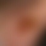
ClinicThis section has been translated automatically.
On pressure or on firm palpation, moderate (about 20%), very severe (60%), to intolerable (20%) pain; spontaneously occurring, rapidly growing, skin-colored or whitish, often centrally ulcerated or encrusted dermal nodule (rarely nodulation), which is usually not displaceable on its base.
About 60% of patients report a very intense stabbing pain, sometimes shooting in spontaneously, which can last minutes but also up to several hours; characteristic is the statement of the patients that they could not sleep on this ear anymore.
The size is usually 0.4-0.7 cm in diameter; the maximum extension is about 2.0 cm in diameter.
CNCH localized atypically (not at the helix) may present as a flat painful ulcer or as a flat keratotic papule or plaque.
HistologyThis section has been translated automatically.
Differential diagnosisThis section has been translated automatically.
Basal cell carcinoma: Absence of pain; ulceration possible; in these cases smooth, "basalcell carcinoma-typical " border.
Gouty tophi: Rarely as a single finding; usually multiple; initially soft, yellowish, later increasingly coarse, usually painless nodules due to deposition of precipitated uric acid salts in the skin.
Keratosis actinica: Usually flat adherent scaling; mild pain may be present, but not intense pain.
Keratoacanthoma: Rapidly growing red nodule, no painfulness.
Squamous cell carcinoma: Important DD; clinically not always definite although the typical pain of CNCH is absent. Histology is conclusive.
TherapyThis section has been translated automatically.
Surgical procedures:
- The safest therapy is the complete surgical removal of the painful nodules by means of wedge excision, since recurrences can only be prevented if the removal is sufficiently extensive.
- A surgical therapy variant is the so-called "punch and graft procedure". In this technique, the lesional skin with the underlying cartilage is punched out and the defect is covered with full thickness skin.
Alternative: Intralesional glucocorticoid injections with triamcinolone (e.g. Volon A crystal suspension diluted 1:5 with LA such as lidocaine 1%). Initial success is good, with prompt diminishing pain. Long-term success is doubtful.
Alternative: therapeutic trial by electrocautery and curettage for superficial changes may be considered.
Alternative: Laser ablation byCO2 or Erbium YAG laser.
Alternative: Topical glucocorticoids (Note: only moderate chance of success in chronic conditions).
Alternative: Pressure relief by individually adapted ear protection bandage. If necessary, pillow with auricular recess ("donut pillow").
LiteratureThis section has been translated automatically.
- Greenbaum SS (1991) The treatment of chondrodermatitis nodularis chronica helicis with injectable collagen. Int J Dermatol 30: 291-294
- Hesse G et al (1994) Argon laser therapy of chondrodermatitis nodularis chronica helicis. dermatologist 45: 222-224
- Khurana U et al (2015) A man with painful nodules on both ears. Chondrodermatitis nodularis chronica helicis. JAMA Otolaryngol Head Neck Surg 141:481-482
- Nadaud B et al (2014) Chondrodermatitis nodularis helicis. Ann Dermatol Venereol 141:306-307
- Oelzner S et al (2003) Bilateral chondrodermatitis nodularis chronica helicis on the free border of the helix in a woman. J Am Acad Dermatol 49: 720-722
- Rajan N et al (2007) The punch and graft technique: a novel method of surgical treatment for chondrodermatits nodularis helicis. Br J Dermatol 157: 744-747
- Taylor MB (1991) Chondrodermatitis nodularis chronica helicis. Successful treatment with the carbon dioxide laser. J Dermatol Surg Oncol 17: 862-864
- Wagner G et al (2011) Clinical forms, differential diagnosis and therapeutic options of chondrodermatitis nodularis chronica helicis Winkler. JDDG 9: 287-291
- Wettlé C et al(2013) Chondrodermatitis nodularis chronica helicis: a descriptive study of 99 patients. Ann Dermatol Venereol 140:687-962
- Winkler M (1915) Chondrodermatitis nodularis chronica helicis. Arch Dermatol Syphilol 121: 278
Recommended articles


Incoming links (9)
Chondrodermatitis nodularis helicis; Collagenosis reactive perforating; Ear knot, elastic; Ear nodules, painful; Elastotic ear knot; Gouty tophi; Otitis; Recurrent polychondritis; Winkler's disease;Outgoing links (7)
Acanthosis; Actinic keratosis; Basal cell carcinoma (overview); Curettage; Gouty tophi; Keratoakanthoma (overview); Laser;Disclaimer
Please ask your physician for a reliable diagnosis. This website is only meant as a reference.
Authors
Last updated on: 21.04.2022
Author: ![]() Prof. Dr. med. Peter Altmeyer
Prof. Dr. med. Peter Altmeyer
Editor: Dr. med. Eva Kämmerer