Image diagnoses for "Plaque (raised surface > 1cm)"
586 results with 2919 images
Results forPlaque (raised surface > 1cm)

Larva migrans B76.9
Larva migrans. linear plaque, subepidermally located, tortuous itchy duct through Ancylostoma brasiliensis on the sole of the foot, existing since about 2 weeks.

Psoriasis (Übersicht) L40.-
Relapsing activity in chronic psoriasis: psoriasis known for a long time. 4 weeks (post-infection) of clear relapsing activity with small papules and plaques. Itching.

Verruciform epidermodysplasia B07.x
Epidermodysplasia verruciformis: disseminated and generalized seeding of flat (planar warts similar to warts); here a section of the forearm
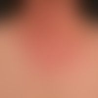
Airborne contact dermatitis L23.8
Airborne Contact Dermatitis: Subacute, blurred, red plaque, here the transition to the non-free skin areas.

Psoriasis capitis L40.8
Psoriasis capitis: chronically inpatient red and white plaques, localised on the forehead and capillitium, reaching far into the hairy area, sharply defined. currently, after insufficient pre-treatment. further red plaques on the elbows.

Circumscribed scleroderma L94.0
Circumscribed scleroderma. Atrophy of the right leg muscles, atrophy of the gluteal muscles on the right, shortening of the right leg (difference 2.0 cm) with consecutive secondary pelvic obliquity and scoliosis in a 19-year-old female patient. The right knee joint is massively restricted in its movement (extension/flexion 0/25/100).

Keratosis palmoplantaris diffusa with mutations in KRT 9 Q82.8
Keratosis palmoplantaris diffusa circumscripta. 2-year-old boy has a chronic, congenital, smooth, evenly distributed, waxy thickened and yellowish discolored plaque formation of both palms. No symptoms. It is an autosomal dominant inherited palmoplantar cornification disorder.

Vaccinations skin changes
Influenza vaccinations, skin changes:initially blistery, later purulent local reaction after influenza vaccination.

Mycosis fungoides C84.0
Mycosis fungoides: tumor stage. 53-year-old man with multiple, disseminated, 1.0-5.0 cm large, in places also large-area, moderately itchy, clearly consistency increased, red, rough, confluent plaques.

Lupus erythematodes chronicus discoides L93.0
Lupus erythematodes chronicus discoides , chronic moderately indurated plaques, marginal with inflammatory activity, central scarring.

Vulvar lichen sclerosus N90.4
Lichen sclerosus of vulva: homogeneous whitish sclerosis of vulva and perineum. 6-year-old girl.

Radiodermatitis chronic L58.1
Radiodermatitis chronica. 72-year-old female patient who was radiated 15 years ago because of a left-sided breast carcinoma. 15 years ago. 15 years ago. 15 years ago. 15 years ago. 15 years ago. 15 years ago. 72-year-old female patient who was radiated because of a left-sided breast carcinoma. 15 years ago. 15 years ago. 15 years ago. 15 years ago. 72-year-old female patient who was radiated because of a left-sided breast carcinoma. 15 years ago. With extensive induration of the skin, a colorful-checked picture with bizarre white spots, flat or linear red spots (telangiectasia) as well as scaling and crust formation over corresponding ulcerations appears.

Cutaneous mastocytoma Q82.2
Mastocytoma kutanes: 1.0 x 2.0 cm, yellow-brown, flat, crescent-shaped, raised lump with blurred edges, protruding in the first two months of life; normal surface relief above the lump.

Psoriasis vulgaris chronic active plaque type L40.0
Psoriasis vulgaris chronic active plaque type: relapsing-active plaque psoriasis.

Keratosis areolae mammae acquisita L 82
Keratosis areolae mammae as side effect of a therapy with vemurafenib (see also there).

Chilblain lupus L93.2
Chilblain lupus - temperature dependent redness, swelling and painfulness of several toes.
