Image diagnoses for "Leg/Foot"
405 results with 1181 images
Results forLeg/Foot

Lupus erythematosus acute-cutaneous L93.1
lupus erythematosus acute-cutaneous: clinical picture known for several years, occurring within 14 days and still with relapsing course at the time of admission. in contrast to the anular pattern on the trunk, irregular, blurred red plaques. in the current relapsing phase fatigue and exhaustion. ANA 1:160; anti-Ro/SSA antibodies positive. DIF: LE - typical.


Lipoid proteinosis E78.8
Hyalinosis cutis et mucosae: psoriasiform, scaly plaques on both knees. No itching.

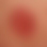
Nummular dermatitis L30.0
Nummular dermatitis: Extensive nummular lesions that havebeen present for several months with blurred, considerably itchy papules and confluent plaques. No hinwesi for psoriasis. No evidence of atopic diathesis.

Pyoderma gangraenosum L88

Keratosis palmoplantaris diffusa with mutation in keratin 1 Q82.8
Keratosis extremitatum hereditaria transgrediens et progrediens


Klippel-trénaunay syndrome Q87.2
Klippel-Trénaunay syndrome: extensive vascular malformation with extensive nevus flammeus affecting the trunk and both arms. So far no evidence of soft tissue hypertrophy. No AV fistulas.


Contact dermatitis toxic L24.-
Toxic contact dermatitis: Enlargement of a section: extensive redness and swelling, in places with confluent formation of vesicles and blisters; beginning scaling (central section).


Acrodermatitis chronica atrophicans L90.4
Acrodermatitis chronica atrophicans. general view: blurred, livid red, spots on the right thigh. skin in the lower area (arrow mark) folded like cigarette paper

Larva migrans B76.9
Larva migrans. Characteristic, itchy, linear gait structures on the sole of the foot.


Idiopathic guttate hypomelanosis L81.5

Prurigo simplex subacuta L28.2
Prurigo simplex subacuta: unusually extensive clinical picture with papules of different sizes, always centrally excoriated in a 51-year-old type I diabetic. severe, always punctual, prickly itching. ?spooning? of the lesion with the fingernail and then sudden cessation of the itching. involvement of the upper arm extensor sides, upper back, thigh extensor sides, chest region and face.

Sarcoidosis of the skin D86.3
Sarcoidosis. chronic sarcoidosis without detectable organ involvement. several to 10.0 cm large, anular, completely symptom-free, brown-red plaques with a smooth surface.