Image diagnoses for "Leg/Foot"
405 results with 1181 images
Results forLeg/Foot


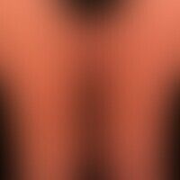

Congestive dermatitis I83.1
stasis dermatitis: flat, sharply limited plaque of the entire right lower leg. lipofasciosclerosis in case of a previously known CVI with beginning papillomatosis cutis lymphostatica. condition after leg ulcer. currently distinct exudation, lymphorrhoea as well as secondary bacterial colonization.

Lichen amyloidosis E85.4
Lichen amyloidosus, detail enlargement: Densely standing, skin-coloured, pinhead-sized, itchy papules on sclerotically altered surroundings.

Larva migrans B76.9
Larva migrans. linear plaque, subepidermally located, winding passage through Ancylostoma brasiliensis in the area of the foot.


Vasculitis (overview) L95.8


Lipoatrophy, localized after glucocorticosteroid injections T88.7
lipoatrophy, localized after glucocorticosteroid injections. general view: 2.5 x 3.0 cm large, circular area with whitish atrophy of the skin and telangiectasia. clear loss of substance of subcutis and fatty tissue. distal of the atrophy a slight swelling in the sense of a lymphatic congestion is visible. the skin changes developed in the course of the last two years, after a single steroid injection into the left knee because of knee problems.


Purpura thrombocytopenic M31.1; M69.61(Thrombozytopenie)
Purpura thrombocytopenic: line shaped (after scratching, as well as after application of a compression bandage) fresh and slightly older skin bleedings (cannot be pushed away diascopically).

Arterial leg ulcer L98.4
Ulcus cruris arteriosum: Sharply defined, painful ulcer on the back of the foot that seizes the tendon.


Acrodermatitis chronica atrophicans L90.4
Acrodermatitis chronica atrophicans. Clearly visible, flaccid skin atrophy and edematous redness on the right foot in a serologically proven infection with Borrelia bacteria. The patient spends several months every summer in the Black Forest.

Asymmetrical nevus flammeus Q82.5
Nevus flammeus: congenital, asymmetrically arranged, non-syndromal (no tissue hypertrophy, no orthopedic malposition) large-area (telangiectatic) vascular nevus; characteristic are the scattered borders of the red spots.


Lymphomatoids papulose C86.6
Lymphomatoid papulosis: chronic, relapsing, completely asymptomatic clinical picture with multiple, 0.3 - 1.2 cm large, flat, scaly papules and nodules as well as ulcers. 35-year-old otherwise healthy man.

Klippel-trénaunay syndrome Q87.2
Klippel-Trénaunay syndrome: extensive vascular malformation with extensive nevus flammeus affecting the trunk, the right arm and both legs. No evidence of soft tissue hypertrophy so far. No AV fistulas.

Metastases C79.8
Metastasis: Multiple, differently sized, partly reddish, partly darkly pigmented smooth nodules on the thigh in patients with malignant melanoma.