Synonym(s)
DefinitionThis section has been translated automatically.
Mostly chronic, superficial, inflammatory or non-inflammatory tinea of the hairless skin with centrifugal spread.
PathogenThis section has been translated automatically.
The frequency of certain pathogens varies in the different continents.
In Europe, Trichophyton rubrum, Trichophyton mentagrophytes, and Epidermophyton floccosum are the most common. See also Dermatophytes. Tinea corporis gladiatores is mostly caused by T. tonsurans.
In a larger Persian study, the pathogen frequency was detected as follows: Trichophyton verrucosum (40.6%), Trichophyton mentagrophytes (17.6%) , Trichophyton violaceum (12%).
In an Indian study, Trichophyton mentagrophytes was found most frequently (63%), followed by Trichophyton rubrum (35%).
You might also be interested in

EtiopathogenesisThis section has been translated automatically.
Frequently infection by domestic animals (cats, cattle, guinea pigs, hamsters - see also zoonoses).
Initially described as folliculitis resulting from the penetration of the fungus into the follicleostium and hair follicles.
The infection spreads exclusively in the stratum corneum with consecutive (optional) infestation of further hair follicles (formation of follicular papules).
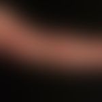
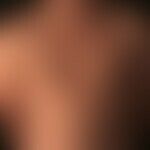
Clinically, an inflammatory, slightly scaly, red, roundish plaque with centrifugal expansion and central healing tendency is observed.
As the infection progresses, several such foci may confluent and form polycyclic, large, map-like figures.
ManifestationThis section has been translated automatically.
Frequently occurring in children, but also in adults.
LocalizationThis section has been translated automatically.
Face(tinea faciei), neck (tinea colli), trunk, extremities (tinea corporis), feet/hands(tinea pedis, tinea manuum), genital area.
ClinicThis section has been translated automatically.
Initially, a circumscribed folliculitis develops as a result of the penetration of the fungus into the follicleostium and hair follicles (clinically rarely observed). The infection spreads exclusively in the stratum corneum and infests other hair follicles.
In general, one or more inflammatory reddened, slightly scaly, border-emphasized plaques with a centrifugal expansion tendency appear at the initial diagnosis. Inflammatory follicular papules are often included.
As the infection progresses, several foci may confluent and form polycyclic, large, map-like figures with central healing.
There is almost always a marked itching, which often leads to pre-treatment with corticosteroids and thus obscures the clinical inflammatory pattern of tinea.
Less frequent is the development of a tinea profunda which is caused by an excessive inflammatory reaction of the organism to the dermatophyenic infection of the skin (analogous to the Kerion Celsi of the capillitium). Clinically impressive are inflammatory, moderately painful, frequently verrucous plaques and nodules which may be interspersed with follicular pustules (see also Granuloma trichophyticum) . Majocchi granuloma is described as granulomatous and pustular tinea caused by Trichophyton rubrum. Nodular abscesses are found mainly in immunocompromised persons.
Under prolonged and severe immunosuppression an erosive and necrotizing tinea corporis is observed, often complicated by a superimposed superinfection by Staphylococcus aureus.
HistologyThis section has been translated automatically.
Mild to moderate acanthosis of the surface epithelium (see Figs. A-B) with focal spongiosis and parakeratosis. In early phases of infection, a rather sparse perivascular lymphocyte infiltrate is found with papillary edema, focal epitheliotropy, and spongiosis to spongiotic vesicles. With prolonged persistence, increasing exocytosis of neutrophil granulocytes, which may condense sub- or intracorneally into small neutrophil abscesses. In the upper dermis, there is a dense diffuse, sometimes perivasally accentuated lymphocytic infiltrate with few neutrophils and eosinophilic granulocytes.
On PAS staining, ( if there was no antifungal pretreatment,) mycelia can be detected at varying densities at the border between the stratified ortho- and parakeratotic zones.
If there is an unusual density of mycelia in the stratum corneum and in the follicular keratin, immunosuppression should be considered.
Differential diagnosisThis section has been translated automatically.
- Clinical Differentials:
- Eczema, nummular: Eczematized plaques without prominent margins; usually disseminated, no mycelia in PAS preparation.
- Seborrheic eczema: No itching; in the case of marginalized plaques, these cannot be differentiated from tinea corporis in terms of differential diagnosis! Typical is the recurrent course of the disease with intensification in the winter months and possibly complete healing under summer, maritime climate. This course completely excludes a tinea. Neither cultural nor histological evidence of mycological pathogens can be found.
- Psoriasis vulgaris: Typical plaque psoriasis as an important DD. The Auspitz phenomenon is always negative in tinea corporis!
- Parapsoriasis en plaques: No marginal accentuation, usually no desquamation. Pseudoatrophy! No itching!
- Mycosis fungoides: No border accentuation; initially little or no itching; histology is diagnostic!
- Erythema anulare centrifugum: Very prominent anular configurations; usually scale-free. Itching is absent or very mild. The marginal zone is very distinctly indurated (wet wool thread). Neither cultural nor histological evidence of mycological pathogens is successful.
- Early syphilis: syphilides are usually accompanied by LK swelling. Distribution pattern: frequent infestation of palms and face. Serology is conclusive! Histology is conclusive (plasma cell dermatitis).
- Erythema migrans: Surface smooth, non-pruritic, marginal plaque. Serology is diagnostic.
- Histologic differential diagnoses (often difficult to distinguish from tinea and can be definitively evaluated only in the context of the clinical picture; therefore, good clinical information is important):
- Acute and subacute eczema: spongiosis, areal parakeratosis, in atopic eczema possible prominent eosinophilia. Often indistinguishable.
- Psoriasis vulgaris: Acanthosis is usually more prominent than in tinea. Areal hyper- and parakeratosis with neutrophil inclusions. The histologic picture is very similar. No evidence of mycelia.
- Parapsoriasis en plaques: fibrosis of the stratum papillare. The surface epithelium is rather atrophic. Epidermotropy is present. Hardly any spongiosis. No parakeratosis!
- Impetigo contagiosa: Subcorneal cleft formation with accumulation of neutrophilic granulocytes. No evidence of mycelium!
- Pityriasis rosea: Nonspecific superficial dermatitis with moderate acanthosis. Sometimes erythrocyte extravasations are seen, which are absent in tinea. No mycelia.
- Allergic contact dermatitis: Clinically clearly distinguishable! Marked, planar spongiosis; acanthotic surface epithelium; long mounds of parakeratosis. Histologically can only be differentiated with certainty in conjunction with clinical data.
- Erythema anulare centrifugum: Histological differentiation can only be made in conjunction with clinical data! Mostly dense perivascular infiltrates.
- Early syphilis: Interface dermatitis with psoriasiform epidermal reaction. Dense, band-like infiltrate in upper and middle dermis (lymphocytes, histiocytes and plasma cells. Extension of the infiltrate to the deep vascular plexus.
TherapyThis section has been translated automatically.
General therapyThis section has been translated automatically.
For extensive mycoses:
Terbinafine systemic: In Switzerland and Austria Terbinafine is approved for oral use in children, in Germany however not yet (off-label use). A careful documentation and education of parents about the off-label use and the effect of Terbinafine in studies should be carried out.
The following dosage recommendations apply: Children with 10-20 kg bw: 62.5 mg/day; 21-40 kg bw: 125 mg/day; > 40 kg bw: 250 mg/day. 2-3 weeks of therapy are usually sufficient.
Alternatively: Itraconazole 100mg p.o./day (Sempera®) over a period of 2-4 weeks.
Alternative: Fluconazole 50mg p.o./day (Diflucan®) over a period of 2-7 weeks.
Progression/forecastThis section has been translated automatically.
NaturopathyThis section has been translated automatically.
AftercareThis section has been translated automatically.
Note(s)This section has been translated automatically.
Occupational disease Dermatomycosis: Dermatophyte infections transmitted from animals to man may be recognised and compensated as occupational diseases in accordance with point 3102 of the list of occupational diseases, provided that a causal link with the occupational activity can be demonstrated (see occupational disease of the skin) Infectious diseases that are transmitted from person to person and affect insured persons who work in the health care system or are exposed to a similar degree to the risk of infection through another activity can be compensated in accordance with BK number 3101.
Fungus detection. In individual cases, the inspection of the entire integument by means of UV light (wood light) has also proved useful for detecting clinically only discretely affected areas with certain dermatophytes or Malassezia species.
Tinea genitalis as venereal disease (especially in tropical zones): Trichophyton interdigitale can be transmitted during sexual intercourse. It can lead to massive infections, especially in patients who shave regularly in the genital area (damage to the epidermal barrier).
LiteratureThis section has been translated automatically.
- Angerstein JH (1977) How tea tree oils heal. With recipes for the most common complaints. Midens publishing house, Augsburg
- Bhatia VK et al. (2014) Epidemiological studies on Dermatophytosis in human patients in Himachal Pradesh, India. Springerplus. 3:134
- Chadeganipour M et al. (2015) A 10-Year Study of Dermatophytoses in Isfahan, Iran. J Clin Lab Anal doi: 10.1002/jcla.21852
- Kassem MA et al. (2007) Efficacy of topical griseofulvin in treatment of tinea corporis. Mycoses 49: 232-235
- Leiva-Salinas M et al. (2014) Tinea capitis in schoolchildren in a rural area in southern Ethiopia. Int J Dermatol 54: 800-805
- Luchsinger I et al. (2015) Tinea genitalis: a new entity of sexually transmitted infection? Case series and review of the literature. Sex Transm Infect 91:493-496.
- Seebacher C et al (2007) Tinea of the free skin. J Dtsch Dermatol Ges 11: 921-926
- Thakur R (2015) Spectrum of dermatophyte infections in Botswana. Clin Cosmet Investig Dermatol 8:127-133
Uhrlass S et al. (2022) Trichophyton indotineae-An Emerging Pathogen Causing Recalcitrant Dermatophytoses in India and Worldwide-A Multidimensional Perspective. J Fungi (Basel) 8:757.
- Wilmer A et al. (1998) Systemic terbinafine treatment of dermatophytoses in children. Mycoses 41: 54-57
- Young Woon Park et al. (2014) Clues' for the Histological Diagnosis of Tinea: How Reliable Are They?
Ann Dermatol 26:286-288
Recommended articles


Incoming links (48)
Amorolfin; Annular dermatoses; Asymmetric periflexural exanthema of childhood ; Basal cell carcinoma superficial; Dartres furfuracees arrondies; Dermatomycoses; Dermatophytosis syndrome; Eccema marginatum (hebra); Epidermophyton floccosum; Erythema; ... Show allOutgoing links (30)
Antimycotics; Atopic dermatitis (overview); Contact dermatitis allergic; Contagious impetigo; Dermatophytes; Early syphilis; Eczema (overview); Epidermophyton floccosum; Erythema anulare centrifugum; Erythema migrans; ... Show allDisclaimer
Please ask your physician for a reliable diagnosis. This website is only meant as a reference.