Synonym(s)
DefinitionThis section has been translated automatically.
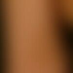
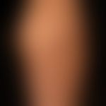
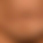
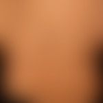
Congenital or acquired in early childhood, homogeneous, pale-brown colored, sharply circumscribed, usually rounded, non-palpable, brown or brown-yellow spots or patches (= spots >1.0 cm ) - see also hyperpigmentation.
Single (up to 3) of these patches are found in 3% of all newborns and in 10-28% of all older children.
If there are 6 or > 6 café au lait spots of > 0.5 cm (prepubertal) and > 1.5 cm (postpubertal), there is evidence of systemic disease. Also in case of very large café au lait spots in segmental or blaschcoid arrangement, a malformation syndrome (see below) must be considered.
Occurrence/EpidemiologyThis section has been translated automatically.
You might also be interested in

ManifestationThis section has been translated automatically.
congenital or acquired in early childhood.
LocalizationThis section has been translated automatically.
Torso and extremities.
ClinicThis section has been translated automatically.
Sharply demarcated, homogeneously colored, circular or oval, non-splashy angular, light brown (milk coffee colored), asymptomatic spots and patches of variable size. Rarer is a systematized arrangement in the Blaschko lines or a strictly hemifacial arrangement (indicative of neurofibromatosis type V). A single very large café-au-lait spot does not appear to indicate a clinical syndrome.
Reported associations of café-au-lait spots with childhood medulloblastomas.
Clinical syndromes with café-au-lait spots (more than 6 café-au-lait spots indicate a syndromal constellation):
- Albright syndrome
- Ataxia teleangiectatica
- Bannayan-Riley-Ruvalcaba syndrome
- Bloom syndrome
- Cobb syndrome (cutaneo-meningo-spinal angiomatosis; associations with segmental neurofibromatosis have been described)
- Cowden syndrome
- Fèvre-Languepin syndrome (popliteal pterygium syndrome, mutation in IRF6 gene)
- Multiple endocrine neoplasia (MEN)
- Neurofibromatosis types I, II, VI
- Noonan syndrome
- Piebaldism
- Tuberous sclerosis
- Turner syndrome.
HistologyThis section has been translated automatically.
S.a. Lentigo simplex. Homogeneous hyperpigmentation of the str. basale (Melan A representation), increased melanocyte numbers, giant melanosomes.
TherapyThis section has been translated automatically.
Usually not necessary, it is rather advised against a therapy. In case of cosmetic indication, possibly an attempt with laser treatment of superficial cryosurgery or dermabrasion. The risk of laser surgery is either recurrence, scarring, depigmentation or a spotty overall result. A study with complete regression through treatment with a pulsed dye laser is available (Goldberg DJ 1997).
Progression/forecastThis section has been translated automatically.
Café au lait stains have no tendency to develop malignancy. Especially during childhood size growth is observed
LiteratureThis section has been translated automatically.
- Goldberg DJ (1997) Laser treatment of pigmented lesions. Dermatol Clin. 15:397
- Kansal NK et al.(2017) Association of piebaldism with café-au-lait macules. Skinmed 15: 223-225.
- Langenbach N et al. (1998) Nevi spili, café-au-lait spots and melanocytic naevi aggregated alongside Blaschko's lines, with a review of segmental melanocytic lesions.Acta Derm Venereol 78:378-380.
- Lo FS et al. (2017) Detection of Rare Somatic GNAS Mutation in McCune-Albright Syndrome Using a Novel Peptide Nucleic Acid Probe in a Single Tube. Molecules 22. pii: E1874.
- Marinău LD et al (2017) Two girl patients with medulloblastoma. Case reports. Rome J Morphol Embryol 58:1103-1108.
- Nguyen JT et al (2004) Large solitary café au lait spots: a report of 5 cases and review of the literature. Cutis 73:311-314
- Shah KN (2010) The diagnostic and clinical significance of café-au-lait macules.Pediatr Clin North Am 57:1131-11253 .
Recommended articles


Incoming links (28)
Ataxia teleangiectatica; Bannayan-riley-ruvalcaba syndrome; Bloom syndrome; Café-au-lait stain; Coast-of-main stains; Cowden syndrome; Fanconi anaemia; Fèvre-languepin syndrome; Hypomelanosis ito; Juvenile xanthogranuloma; ... Show allOutgoing links (25)
Ataxia teleangiectatica; Bannayan-riley-ruvalcaba syndrome; Becker's nevus; Berloque dermatitis; Bloom syndrome; Café-au-lait stain; Cobb syndrome; Cowden syndrome; Cryosurgery; Dermabrasion; ... Show allDisclaimer
Please ask your physician for a reliable diagnosis. This website is only meant as a reference.
Authors
Last updated on: 02.03.2023
Author: ![]() Prof. Dr. med. Peter Altmeyer
Prof. Dr. med. Peter Altmeyer
Editor: ![]() Dr. med. Lucian Cajacob
Dr. med. Lucian Cajacob