Image diagnoses for "Plaque (raised surface > 1cm)", "Leg/Foot"
181 results with 457 images
Results forPlaque (raised surface > 1cm)Leg/Foot
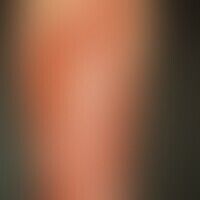
Necrobiosis lipoidica L92.1
Necrobiosis lipoidica: Waxy, reddish-brown, smooth, shiny infiltrate plates with several punched-out ulcers (after banal trauma) in type I diabetes in the area of the tibia.
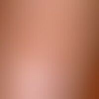
Keratosis seborrhoic (plaque type)
Keratosis seborrhoeic (plaque type): flat, regularly bordered, little pigmented, non-irritant plaque.

Psoriasis palmaris et plantaris (plaque type) L40.3
Psoriasis palmaris et plantaris (plaque-type chronic inpatient plaquepsoriasis of the sole of the foot with coarse desquamation and painful hare formation. no topical pre-treatment

Eosinophilic granulomatosis with polyangiitis M30.1
Churg-Strauss syndrome: encircled central granulomatous regression zone (see histological preparation 1); on the right side the progression zone is outlined (see histological preparation 2).
Necrobiosis lipoidica L92.1
Necrobiosis lipoidica: Overview of the left thigh: Approx. 3 cm large, slightly elevated, erythematous plaque without ulcerations.
Tinea pedis (overview) B35.30
Tinea pedum (moccasin type). general view: For about 13 years non-healing redness and scaling, partly with severe itching, in the area of the right foot in a 30-year-old female patient. sharply defined, marginal scaling erythema, pustular formation, swelling of the foot with limited walking ability.
Tinea pedis (overview) B35.30
Tinea pedum, detail enlargement: Sharply defined, marginal scaly erythema, pustular formation, scaly seam along the edge of the foot and multiple scratch excoriations, some of which are crusty.

Tinea pedis (overview) B35.30
Tinea pedum. general view: Persistent redness and scaling, partly with severe itching, in the area of the left foot in a 30-year-old female patient, which has not healed for about 13 years. sharply defined, marginal scaling erythema, pustular formation.
Varicella B01.9
Varicella: generalized exanthema (aspects of erythema multiforme) with coexistence of larger and smaller papules, vesicles, plaques.
Necrobiosis lipoidica L92.1
Necrobiosis lipoidica: irregularly configured, sharply defined, plate-like, atrophic, "scleroderma-like", smooth plaques. brownish-yellow sunken centre with atrophy of skin and fatty tissue. reddish-violet to brownish-red rim.
Nodular vasculitis A18.4
erythema induratum. solitary, chronically stationary, 4.0 x 3.0 cm in size, only imperceptibly growing, firm, moderately painful, reddish-brown, flatly raised, rough, scaly nodules with a deep-seated part (iceberg phenomenon). intermediate painful ulcer formation (Fig). no evidence of mycobacteriosis.
Eosinophilic cellulitis L98.3
Cellulitis eosinophil: acute formation of circumscribed, large, sharply margined plaques The surface of the plaques may have an orange peel-like texture (see following figure)
Acrodermatitis chronica atrophicans L90.4
Acrodermatitis chronica atrophicans: Initially flat, oedematous, livid red plaques; beginning transition to pronounced, flaccid atrophy with typical wrinkling of the skin (cigarette-paper phenomenon) and clearly translucent vein networks.

Nevus melanocytic acral D22.L
nevus melanocytic acral: completely sympotmless, congenital melanocytic, nevus that covers the sole and back of the foot. bizarre lateral borders, different shades of brown and black. six-monthly controls are indicated.
