Nodular vasculitis A18.4
Erythema induratum (Nodular vasculitis): The 48-year-old patient has been suffering for 2 years from these intermittent, moderately painful, therapy-resistant plaques which tend to ulceration.

Purpura eczematid-like purpura L81.7
Purpura eczematide-like purpura: non-symptomatic (no itching) eczema-like disease that has been recurrent for months in a completely healthy patient (no history of medication).

Nevus pigmentosus et pilosus D22.L6

Granuloma anulare disseminatum L92.0
Granuloma anulare disseminatum: non-painful, non-itching, disseminated, large-area plaques that appeared on the trunk and extremities of a 52-year-old patient. No diabetes mellitus. No other systemic diseases known.

Acquired progressive lymphangioma D18.10
Lymphangioma progressive: large brownish-red plaques, which fray into small flat plaques at the edges. No complaints. We aregratefulto Dr. U. Ammanfor submitting this image.

Scleroderma linear L94.1
Scleroderma ligamentous: for years slowly progressive, only moderately indurated ligamentous morphea in a 42-year-old woman; no movement restrictions of the joints.

Nevus melanocytic congenital D22.-
Nevus, melanocytic, congenital. 6 x 4 mm large, brownish pigmented nevus in the area of the left small toe in a 3-month-old girl. Regular clinical control is necessary. Excision planned at the age of >10 years.

Sarcoidosis of the skin D86.3
Sarcoidosis, plaque form: slightly pressure-painful plaques of the skin with plates with a scaly surface that can be easily distinguished from the surrounding area and can be moved on the support.

Necrobiosis lipoidica L92.1
Necrobiosis lipoidica: Waxy, reddish-brown, smooth, shiny infiltrate plates with several punched-out ulcers (after banal trauma) in type I diabetes in the area of the tibia.
Keratosis seborrhoic (plaque type)
Keratosis seborrhoeic (plaque type): flat, regularly bordered, little pigmented, non-irritant plaque.

Nevus melanocytic congenital D22.-
Nevus melanocytic congenital: large, congenital, hairy melanocytic nevus. No changes during the annual clinical controls. Inlet: reflected light microscopy.

Necrobiosis lipoidica L92.1
Necrobiosis lipoidica: chronic, sharply defined, flat, centrally atrophic, smooth plaque with clearly brown-red tinged edges; shining through of the underlying veins is characteristic.
Purpura pigmentosa progressive L81.7
Purpura pigmentosa progressiva. irregularly configured, reddish-brownish spots with petechiae that cannot be pushed away (cayenne pepper spots).
Circumscribed scleroderma L94.0
Circumscripts of scleroderma (small-heart plaque or confetti type): disseminated, symptomless, 0.1-0.2 cm large, confetti-like, white spots/papules with (incident light microscopic) detectable, atropically shiny surface. The skin lesions have now been discovered (by chance) after sunbathing. Histology: No evidence of Lichen sclerosus.
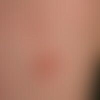
Necrobiosis lipoidica L92.1
Necrobiosis lipoidica: Overview of the left thigh: Approx. 3 cm large, slightly elevated, erythematous plaque without ulcerations.
Purpura pigmentosa progressive L81.7
Purpura pigmentosa progressiva: aetiologically unexplained (medication?) pronounced clinical picture that has been changing for several months with symmetrically distributed, disseminated, non-itching, yellow-brown, spots (detailed picture).

Purpura jaune d'ocre L81.9
Purpura jaune d'ocre: multiple, chronically stationary, proximally disseminated and blurred, distally confluent, symptom-free, light to dark brown, rough, scaly spots of varying intensity, located on the distal lower legs; detectable chronic venous insufficiency (CVI).
Purpura pigmentosa progressive L81.7
Purpura pigmentosa progressiva. incident light microscopy, blurred, yellow-brownish spots (star), in addition to punctiform, fresh bleeding (horizontal arrow) also older brown-reddish spots already in decomposition (vertical arrow). line pattern: traced skin line pattern of the skin of the lower leg

Necrobiosis lipoidica L92.1
Necrobiosis lipoidica: irregularly configured, sharply defined, plate-like, atrophic, "scleroderma-like", smooth plaques. brownish-yellow sunken centre with atrophy of skin and fatty tissue. reddish-violet to brownish-red rim.


