Synonym(s)
HistoryThis section has been translated automatically.
DefinitionThis section has been translated automatically.
Rare, highly malignant and aggressively growing vascular tumour in the sun-damaged scalp of elderly people. It is characterized by rapid and often uncontrollable growth with a very poor prognosis.
You might also be interested in
Occurrence/EpidemiologyThis section has been translated automatically.
m>w; usually 60th to 80th year of life. Angiosarcomas account for about 0.01% of all malignant tumors of the adult and 5% of cutaneous sarcomas.
ManifestationThis section has been translated automatically.
Mostly occurring between 60 and 80 years of age (average age 65 years).
LocalizationThis section has been translated automatically.
ClinicThis section has been translated automatically.
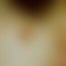
Initially, clinically inconspicuous, locally persistent, blurred, 1.0-3.0 cm large, painless, smooth, telangiectatic red spots appear, which are often misjudged as "banal telangiectasias in the context of rosacea or light-damaged skin" in an early phase of tumor development.
Over the course of months, the area expands; the red coloration of the skin continues to become increasingly intense; these tend to form extensive hematomas. These extensive skin hemorrhages (occurring with banal trauma) should, if other explanations are not valid, lead to the diagnosis of "angiosarcoma of the scalp". Later formation of flat, blue-red papules, plaques and nodules, which tend to ulcerate with the formation of extensive erosions and ulcers. Subsequent lymphorrhea.
Note on clinical differential diagnosis (head-tilt maneuver: filling and becoming visible after a short head-down position (head-tilt maneuver = 20 sec. head-down position) is an important diagnostic sign.
Metastasis: Hematogenous, especially in the lungs and skeletal system. Lymphogenous, in regional lymph nodes.
Note! Always think of angiosarcoma in the case of unexplained extensive hemorrhages in the area of the scalp!
HistologyThis section has been translated automatically.
Initial: Enlarged endothelia lined by atypical, irregularly dilated, bizarre capillary-like structures that protrude into the lumina like buttons and often anastomose with each other. The endothelia show enlarged chromatin-rich nuclei and increased Ki-67 positivity.
Later: Solid masses of polymorphic spindle cells, blood-filled clefts and erythrocyte extravasations.
Immunohistology: Tumor cells express CD31, CD34 and vimentin. The neoplastic vascular spaces are either not or only slightly surrounded by actin-positive cells.
DiagnosisThis section has been translated automatically.
Differential diagnosisThis section has been translated automatically.
- Erythematous rosacea: Mostly bilateral; characteristic concomitant symptoms of rosacea (variable redness with flushing symptoms due to temperature changes, alcohol consumption, etc.; never smudging).
- Morbihan's disease: Mostly bilateral; characteristic accompanying symptoms of rosacea possible. No smugillations.
- Nevus flammeus: History excludes angiosarcoma.
- Hematoma (initial): History excludes angiosarcoma.
- Systemic lupus erythematosus: Symmetry of erythema; disorders of AZ; serologic autoimmune phenomena.
- Pellagroid: Only occurring in exposed areas, itching or burning after UV exposure, color rather red-brown.
- Kaposi's sarcoma: Exclusion of HIV infection (laboratory).
- Sarcoidosis: Chronic persistent red patches or plaques, livid discoloration possible in cold weather, may be associated with lupus pernio. Diascopic: autoinfiltrate. Histologically: Evidence of sarcoid granulomas!
Radiation therapyThis section has been translated automatically.
Larger studies have demonstrated the value of postoperative adjuvant radiotherapy (linear accelerators are recommended) (significant reduction in mortality). Dose: 55-60 Gy, with suspected residual tumor up to 75 Gy. A large safety distance should be maintained. Radiation therapy alone is not recommended, as the clinical results are inferior to combination therapy.
Caution! Recurrences are increasingly less sensitive to radiation!
Internal therapyThis section has been translated automatically.
For non-resectable findings, there is good experience with pegylated liposomal doxorubicin and paclitaxel (Taxol). As a rule of thumb, around 25% of patients respond to doxorubicin-based regimens (50 mg/m2 every 28 days), with the additional administration of ifosfamide achieving a trend improvement, albeit with a significant increase in toxicity.
In a large retrospective analysis (125 patients), the combination of liposomal doxorubicin and paclitaxel was found to be relatively effective, as was the combination of doxorubicin and ifosfamide.
Other possible chemotherapeutic agents for angiosarcoma are docetaxel, vinorelbine, gemcitabine and epirubicin.
Combined chemotherapy/radiotherapy (taxanes) does not appear to achieve any improvement over chemotherapy alone.
Operative therapieThis section has been translated automatically.
Progression/forecastThis section has been translated automatically.
Very poor prognosis for tumours > 4 cm in diameter, as early haematogenic metastasis (lung) occurs. 5-year survival rate is <10%. A better prognosis can be expected for angiosarcomas <4 cm.
Note(s)This section has been translated automatically.
LiteratureThis section has been translated automatically.
- Bong AB et al (2004) Treatment of scalp angiosarcoma by controlled perfusion of a carotis externa with pegylated liposomal doxorubicin and intralesional application of pegylated interferon alpha. J Am Acad 52: S20-S23
- Bork K et al (1985) Undifferentiated cutaneous angiosarcoma of the head. Dermatologist 36: 341-346
- Dhanasekar P et al (2012) Cutaneous angiosarcoma of the scalp masquerading as a squamous cell carcinoma: case report and literature review. J Cutan Med Surg 16:187-190.
- Gonzalez MJ et al (2009) Angiosarcoma of the scalp: a case report and review of current and novel therapeutic regimens. Dermatol Surge 35: 679-684.
- Holden CA et al (1987) Angiosarcoma of the face and scalp, prognosis and treatment. Cancer 59: 1046-1057
- Ito T et al (2016) Cutaneous angiosarcoma of the head and face: a single-center analysis of treatment outcomes in 43 patients in Japan. J Cancer Res Clin Oncol. PMID: 27015673
- Mentzel T (2011) Sarcomas of the skin. Clinics in Dermatology 29: 80-90
- Nishiwaki Y et al (2002) A case of angiosarcoma of the nose. J Dermatol 29: 593-598
- Rosai J et al (1976) Angiosarcoma of the Skin. A clinicopathologic and fine structural study. Human Pathol 7: 83-109
- Sharma S (2012) Angiosarcoma of the scalp associated with Xeroderma pigmentosum. Indian J Med Paediatr Oncol 33:126-129.
Recommended articles


Incoming links (11)
Cutaneous sarcomas (overview); Facial edema; Hemangioendothelioma of the scalp malignant; Hemangiosarcoma of the head and face skin; Idiopathic angiosarcoma; Intravascular papillary endothelial hyperplasia; Lymphangiosarcoma of the scalp; Malignant haemangioendothelioma; Sarcoma angioblastic of the head rind; Targetoid hemosiderotic hemangioma; ... Show allOutgoing links (12)
Doxorubicin; Excision; Gemcitabine; Hematoma; Kaposi's sarcoma (overview); Lupus erythematosus systemic; Lupus pernio; Morbus Morbihan ; Pellagroid; Rosacea; ... Show allDisclaimer
Please ask your physician for a reliable diagnosis. This website is only meant as a reference.


