Lupus erythematosus acute-cutaneous L93.1
lupus erythematosus acute-cutaneous: clinical picture known for several years, occurring within 14 days, at the time of admission still with intermittent course. anular pattern. in the current intermittent phase fatigue and exhaustion. ANA 1:160; anti-Ro/SSA antibodies positive. DIF: LE - typical.


Teleangiectasia macularis eruptiva perstans Q82.2
Teleangiectasia macularis eruptiva perstans. 54-year-old patient with a generalized macular clinical picture which has existed for years and shows a constant progression. Itching in case of heat exposure and mechanical exposure of the affected areas.

Acrodermatitis chronica atrophicans L90.4
acrodermatitis chronica atrophicans. 57-year-old female patient. skin changes existing for five years now, clearly increasing for half a year, discoloured red-blueish in cold weather. large, blurredly limited, symmetrical erythema (completely anaemic). the skin surface is wrinkled in the breast area (atrophic), otherwise smooth. some splatter-like white discolorations.

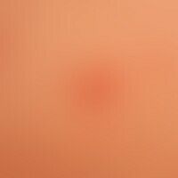
Folliculitis perforating L73.8
Folliculitis, perforating. irregular distribution of follicular, itchy papules with a central horn plug in the area of the back.


Steroid acne L70.8

Cherry angioma D18.01
Angioma senile. red brown, very soft papules, almost completely compressible by finger pressure, 0.7 cm in size. therapy not necessary


Drug effect adverse drug reactions (overview) L27.0

Pyogenic granuloma L98.0
Granuloma pyogenicum (pyogenic granuloma) Rapidly growing, bluish-black, soft, slightly bleeding tumour. Remark: the black colour was caused by thrombosis in the tumour parenchyma.

Sarcoidosis of the skin D86.3
Sarcoidosis: anular or circulatory chronic sarcoidosis of the skin. persisting for several years. onset with small symptomless papules with continuous appositional growth and central healing. no detectable systemic involvement.

Erythema anulare centrifugum L53.1
Erythema anulare centrifugum: Characteristic single cell lesion with peripherally progressive plaque, which flattens centrally and is only recognizable here as a non raised red spot.

Inverted psoriasis L40.83
Psoriasis inversa: 69-year-old woman. 6 months at presentation. no manifestations of psoriasis present on the remaining integument. family history but positive: son with known psoriasis vulgaris.

Zoster B02.9
Zoster: in segmental distribution (Th4), grouped vesicles on reddened skin in a 38-year-old man. Moderate pain. Healing without complications. No postzosteric neuralgia. Here is a detailed picture with fresh grouped vesicles.

Nevus verrucosus Q82.5
Bilateral naevus verrucosus in an infant. No symptoms. Psoriasiform aspect of the plaques running in the Blaschko lines, scattered, reddish, slightly infiltrated, scaly.


Artifacts L98.1

Syphilide papular A51.3


Keloid (overview) L91.0
keloid. large, brown to brown-red, very rough, smooth nodes with a jagged edge structure. not painful to the touch, with significant pressure considerable pain. postoperative condition after excision of several acne nodes in the sternal region.