Image diagnoses for "Plaque (raised surface > 1cm)", "red"
441 results with
1925 images
Results for Plaque (raised surface > 1cm) red
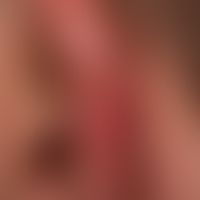
Lupus erythematodes chronicus discoides L93.0
Lupus erythematodes chronicus discoides: blurred, red and brown, partly scaly and crusty, hypersensitive plaque.
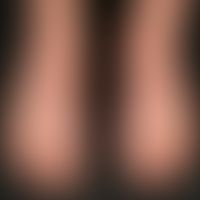
Lichen planus classic type L43.-
Lichen planus (classic type): moderately itchy, disseminated, like scattered distribution pattern, red-violet colour of the surface smooth, shiny papules and plaques.
Lupus erythematosus systemic M32.9
Systemic lupus erythematosus (late onset): chronic, blurred reddish-livid plaques with spatter-like whitish spots, small erosions and crusts; accompanying recurrent fever attacks, fatigue and tiredness, arthralgia, inflammation parameters +, ANA high titer positive, rheumatoid factor +, DNA-Ak+.



Atopic dermatitis (overview) L20.-
Eczema, atopic. chronic, recurrent itchy red spots and slightly raised, flat, rough red plaques on the back of the left hand, the back and the side edges of the fingers of an 8-month-old girl. Furthermore multiple, disseminated, partly crusty scratch excoriations and isolated rhagades are visible.
Lupus erythematodes chronicus discoides L93.0
lupus erythematodes chronicus discoides: 13-year-old otherwise healthy patient. skin lesions since 6 months, gradually increasing, no photosensitivity. several, centrofacially localized, chronically stationary, touch-sensitive (slight pain when stroking with a wooden spatula), red, slightly scaly plaques. histology and DIF are typical for erythematodes. ANA and ENA negative.

Dermatomyositis (overview) M33.-
dermatomyositis. red-violet, slightly itchy, flat. blurred erythema in the décolleté and on the lateral parts of the neck. general fatigue and muscle weakness.
Atopic dermatitis in children and adolescents L20.8
Eczema atopic in childhood: 12-year-old adolescent with generalized atopic eczema. conspicuous grey-brown, dry skin. keratosis pilaris-like follicular keratoses. multiple scratched papules and plaques.
Psoriasis vulgaris L40.00
Psoriasis vulgaris. abbortive form with infestation of the nostrils on both sides. The clinical picture is clinically relevant in that rhagades and pain occur. Local therapy is laborious.

Anticonvulsant hypersensitivity syndrome T88.7

Vulvitis, a-streptococcal vulvitis N76.-
Recurrent perianal dermatitis and vulvitis caused by A-streptococci. 36 year old patient.Fig. from Eiko E. Petersen, Colour Atlas of Vulva Diseases. With the prior approval of Kaymogyn GmbH Freiburg.
Seborrheic dermatitis of adults L21.9
Dermatitis, seborrheic: Blurred, delicately reddened, coarse lamellar scaling, flat, slightly infiltrated plaques in a 44-year-old patient.

Dyskeratosis follicularis Q82.8
Dyskeratosis follicularis. reflected light microscopy: section of a lesion on the neck. yellowish-white keratin plaques (orthohyperkeratosis) and areas with ball-shaped, ectatic central capillaries (acantholysis area).