Image diagnoses for "red"
901 results with 4549 images
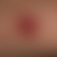
Results forred

Nail hematoma T14.05
Nail hematoma: Apparently caused by repetitive trauma (probably triggered by a trauma from frontal trauma, e.g. during a football match), transverse bleeding, the growing nail area is normally stained.


Gout M10.0
Arthritis urica: sudden spontaneous and painful (difficult to walk) redness and swelling (circled in black) of the right metatarsophalangeal joint of the big toe.


Plaques muqueuses A51.32
Plaques muqueuses: disseminated, localised red, symptom-free plaques confluent in the centre of the tongue with preserved papilla structure (see following figure).

Rhagade R23.4
Rhagade in chronic actinic cheilitis: A deep, narrow, elongated defect of the lower lip which has existed for several weeks and is covered with crusts.


Lupus erythematodes chronicus discoides L93.0
Lupus erythematodes chronicus discoides. overview: Disseminated, scarring, leading to the loss of hair follicles (clinically scarring alopecia), high parietal in a 52-year-old. surrounded by still active foci of CDLE. in the square a scarring area with in places still preserved, in places already destroyed (arrow mark) follicular structure.

Vasculitis leukocytoclastic (non-iga-associated) D69.0; M31.0
Vasculitis leukocytoclastic (non-IgA-associated): multiple, since about 1 week existing, localized on both lower legs, irregularly distributed, 0.1-0.2 cm large, confluent in places, symptomless, red, smooth spots (not compressible).

Demodex folliculitis B88.0
Demodex folliculitis: chronic bilateral follicular dermatitis with extensive reddening. previously known rosacea. for months, however, unexpected significant worsening of the findings. S following figure.


Malasseziafolliculitis B36.8
Malasseziafolliculitis: disseminated, follicle-bound, inflammatory, 0.5-3 mm papules and papulopustules on the back of a 32-year-old female patient; frequent, even long-term, antibiotic therapy due to bacterial cystitis.


Ecchymosis syndrome, painful R23.8
Ecchymosis syndrome, painful seti 6 months of recurrent, painful, extensive skin bleeding on the abodes and extremities in an otherwise healthy 69-year-old female patient

Asymmetrical nevus flammeus Q82.5
Naevus flammeus lateralis. congenital, generalized, spotty erythema on the left arm in an 18-month-old boy with age-appropriate development.

Boils L02.92
Boils. 38-year-old patient with recurrent boils. 2.5 x 2.0 cm large, spontaneously formed, pressure-marked, flat raised lump with yellow necrotic tip and scaly crown.

Leishmaniasis (overview) B55.-
Leishmaniasis (mucocutaneous leishmaniasis of the old world): Ethiopian (HIV-infected) patient with skin and mucous membrane infestation