Synonym(s)
black dots; black points; Brown/black dots on a blue/grey background; brown dots; peripheral black dots; pigment dots
DefinitionThis section has been translated automatically.
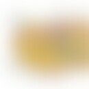
Reflected light microscopic phenomenon caused by punctiform accumulations of melanin pigment in the stratum corneum (black) or in the upper epidermis (brown), measuring about 0.05-0.15 mm in horizontal diameter. The pigment is found both within melanocytes and extracellularly.
General informationThis section has been translated automatically.
Reflected light microscopy: Dark brown or black round, oval or polygonal disc-shaped pigment densities are distributed irregularly in the epidermis either solitarily or multiple times. The spots are located in front of a blue, blue-grey or grey background (melanophagus agglomerates or deep melanocyte nests).
You might also be interested in
OccurrenceThis section has been translated automatically.
If the pigment dots or spots are located in front of a more or less bluish coloured background, the specificity for malignant melanomas within the group of pigment cell tumours is above 80% (sensitivity 47%). An irregular distribution of dots in the tumor periphery is also an indication of the presence of a malignant melanoma or a dysplastic nevus. In other melanocytic nevi, such as spindle cell nevi, congenital nevi or junctional nevi, the dots are usually evenly distributed over the lesion and the background pigment rarely assumes a bluish colour.
HistologyThis section has been translated automatically.
Histologically, superimposed melanin pigment can be detected. In the stratum corneum melanin appears black, in keratinocytes, epidermal naevocytes or melanocytes it is black to dark brown, in the basal cell layer and junctional zone it is reddish brown, brown or light brown, in the upper stratum papillary and the papilla tips grey, grey-brown or grey-blue, in the deep stratum papillary, at the border to the stratum reticulare, steel blue to turquoise. Melanin pigment incorporated by corial melanophages appears grey, grey-blue, grey-violet or steel-grey, depending on the depth of localisation.
LiteratureThis section has been translated automatically.
- Bahmer FA, Fritsch P, Kreusch J, Pehamberger H, Rohrer C, Schindera I, Smolle J, Soyer HP, Stolz W (1990) Diagnostic criteria in reflected light microscopy. Dermatologist 41: 513-514
- Ehring F, Schumann J, Voss W (1977) Vital microscopy in reflected light. Research Reports of the State of NRW No. 2621 Westdeutscher Verlag
- Kenet RO, Kang S, Kenet BJ, Fitzpatrick TB, Sober AJ, Barnhill RL (1993) Clinical diagnosis of pigmented lesions using digital epiluminescence microscopy. Arch Dermatol 129: 157-174
- Rhodes AR (1995) Comment on: "Malignant melanoma in epiluminescent microscopy". In: Sober AJ, Fitzpatrick TB (eds) Yearbook of Dermatlogy 1995, Mosby, St.Louis Baltimore Boston
- Schulz H (2002) Reflected light microscopic vital histology. Springer, Berlin Heidelberg New York
- Yadav S, Vossaert KA, Head AW, Silverman M, Grin-Jorgensen C (1993) Histopathologic correlates of structures seen on dermoscopy (epiluminescence microscopy). On J Dermatopathol 15: 297-305