DefinitionThis section has been translated automatically.
Most frequent neurogenic tumour, which occurs solitary or as part of neurofibromatosis .
ClassificationThis section has been translated automatically.
A distinction is made between:
- Diffuse (superficially located) neurofibroma
- Plexiform (encapsulated, usually deep dermal or subcutaneous) neurofibroma
- Pigmented neurofibroma (diffuse or plexiform)
- Mixed neurofibroma (diffuse and plexiform).
You might also be interested in

ClinicThis section has been translated automatically.
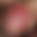
Solitary or multiple, 0.2-0.5 cm large or monstrous, saccular-polyploid, skin-colored to bluish, broad or pedunculated, strikingly soft tumors, possibly with wrinkling or as a dewlap-like structure.
Typical is the bell-button phenomenon: tumors protuberating the skin can be pushed back with a finger through a hernia-like dermal gap. The plexiform (encapsulated) neurofibromas can be palpated as cord-like, coarse structures in the depth of the dermis or subcutis.
HistologyThis section has been translated automatically.
Cell-rich, almost homogeneous accumulation of myelinated and non-myelinated nerve fibres, modified Schwann cells, fibroblasts, macrophages, mast cells, collagenous and elastic fibres and weakly basophilic mucoidal basic substance without recognisable cell boundaries.
Differential diagnosisThis section has been translated automatically.
TherapyThis section has been translated automatically.
In case of functionally or cosmetically disturbing tumours, excision if necessary.
LiteratureThis section has been translated automatically.
- Babovic S et al (2003) Liposuction: a less invasive surgical method of debulking plexiform neurofibromas. Dermatol Surgery 29: 785-787
- Filippo G et al (1989) Solitary Plexiform Neurofibroma. Dermatologica 179: 84-86
- Renshaw A et al (2003) Massive plexiform neurofibroma with associated meningo-encephalocoele and occipital bone defect presenting as a cervical mass. Br J Plast Surgery 56: 514-517
Recommended articles


Incoming links (14)
Bell button phenomenon; Cutis verticis gyrata; Fibrokeratome acquired digital; Gardner syndrome; Legius-syndrome; Molluscum fibrosum; Naevus melanocytic common; Neural tumors; Neurofibroma, pigmented; Neurofibromatosis noonan syndrome; ... Show allOutgoing links (6)
Dermatofibroma; Excision; Neurofibromatosis (overview); Neurom kutanes (overview); Nevus melanocytic (overview); Schwannom;Disclaimer
Please ask your physician for a reliable diagnosis. This website is only meant as a reference.