Synonym(s)
DefinitionThis section has been translated automatically.
Acquired, roundish, oval or also irregularly configured, light to dark brown, usually sharply delimited spot(s) in chronically light-exposed skin areas. Lentigo solaris is caused by an initial purely melanocytic proliferation, which, if prolonged, may be followed by a mixed epidermo-melanocytic proliferation. Lentigo solaris may, depending on the predominant proliferation pattern, change into:
- a verruca seborrhoica of the reticular-acanthotic type
- a melanocytic nevus of the junctional type
- a lentigo maligna
Only lentigo maligna requires treatment.
Occurrence/EpidemiologyThis section has been translated automatically.
In the Central European population, 90% of the 60-year-olds are present in one or more cases. No gender prevalence.
You might also be interested in

EtiopathogenesisThis section has been translated automatically.
Chronic UV exposure.
ManifestationThis section has been translated automatically.
Occurs in the middle (40-60 years) and higher (> 60 years) age groups. No gender preference.
LocalizationThis section has been translated automatically.
Especially face, back of the hand, forearm extensor sides. In women, lower legs and the décolleté are also frequently affected. In fair-skinned (genuinely redheaded) persons, multiple lentigines are also found on the shoulders and back.
ClinicThis section has been translated automatically.
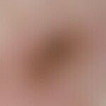
- A few millimetres to several centimetres in size, oval or roundish, also bizarrely configured, sharply defined, yellow-brown to brown spot. Increase in pigmentation due to exposure to sunlight. Only slight fading in winter. Multiple lentigines as they occur after excessive and recurrent UV exposure of the skin are called solar lentiginosis.
- Reflected light microscopy: Regular pigment network with varying degrees of pigmentation.
HistologyThis section has been translated automatically.
Differential diagnosisThis section has been translated automatically.
Verruca seborrhoica; Ephelides; Lentigo maligna; Non-solar lentiginosis may be symptomatic of syndromal disease. See below Lentigines syndromes, pigmented actinic keratosis.
TherapyThis section has been translated automatically.
- Symptomatic; cosmetic coverage with commercial make-up.
- Initially, an experiment with a 5% hydroquinone ointment can be carried out under controlled application. S.a. the application of Resorcin.
- Superficial curettage with sharp curette (Fa. Stiefel).
- Cryosurgery (cryopeeling); in an open spray procedure short "freezing" of the lentiginous lesion.
- In any case, for prophylactic reasons, light protection (especially postoperatively), either textile or with commercially available preparations (e.g. Eucerin Sun, Anthelios).
Progression/forecastThis section has been translated automatically.
NaturopathyThis section has been translated automatically.
LiteratureThis section has been translated automatically.
- Andersen WK et al (1997) Histopathology of solar lentigines of the face: a quantitative study. J Am Acad Dermatol 36: 444-447
Bottoni U et al (2013) Ink spot lentigo: singular clinical features in a
caseseries of patients. Int J Immunopathol Pharmacol 26:953-955- Bastiaens MT et al (1999) Ephelides are more related to pigmentary constitutional host factors than solar lentigines. Pigment Cell Res 12: 316-322
- Kang S et al (2003) Assessment of adapalene gel for the treatment of actinic keratoses and lentigines: a randomized trial. J Am Acad Dermatol 49: 83-90
- Kawada A et al (2002) Videomicroscopic and histopathological investigation of intense pulsed light therapy for solar lentigines. J Dermatol Sci 29: 91-96
- Kawada A et al (2002) Clinical improvement of solar lentigines and ephelides with an intense pulsed light source. Dermatol Surge 28: 504-508
Nakamura M et al (2015) Environment-Induced Lentigines (EILs): Formation of solar lentigines beyond ultraviolet radiation. Exp Dermatol doi: 10.1111/exd.12690
Praetorius C et al (2014) Sun-induced freckling: ephelides and
- solar lentigines. Pigment Cell Melanoma Res 27:339-350
- Todd MM et al (2000) A comparison of 3 lasers and liquid nitrogen in the treatment of solar lentigines: a randomized, controlled, comparative trial. Arch Dermatol 136: 841-846
Recommended articles


Incoming links (14)
Age pigmentation; Age spot; Albinism oculocutaneous tyrosinase-positive; Atrophy senile of the skin; Black ink spot; Flash lamps; Keratosis benign lichenoid; Keratosis seborrheic verruca-plana-like; Lentigo maligna; Lentigo simplex; ... Show allOutgoing links (11)
Cryosurgery; Curettage; Ephelids; Hydroquinone ointment 3%; Keratosis seborrhoeic (overview); Lentigines syndromes; Lentiginosis; Lentigo maligna; Light protection; Resorcin; ... Show allDisclaimer
Please ask your physician for a reliable diagnosis. This website is only meant as a reference.