Synonym(s)
HistoryThis section has been translated automatically.
Hoffmann, 1909; Gougerot and Burnier, 1930
DefinitionThis section has been translated automatically.
Characterized by indurated, surface-smooth, red papules and plaques, non-scarring (from lat tumidus: puffy) variant of cutaneous lupus erythematosus, without significant epidermal component such as scaling or follicular hyperkeratosis. There are smooth transitions to lupus erythematosus profundus. Clinically prominent is a high photosensitivity.
You might also be interested in

Occurrence/EpidemiologyThis section has been translated automatically.
Occurs in about 15% of cases of cutaneous lupus erythematosus.
ManifestationThis section has been translated automatically.
Especially in young adulthood. Age peak: 30th-40th LJ. Less common in children. No sex preference.
LocalizationThis section has been translated automatically.
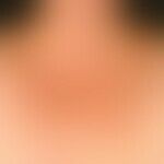
Preferably on the face; neck, décolleté, arms, shoulders may also be affected.
Clinical featuresThis section has been translated automatically.
At light-exposed sites, localized or also disseminated, chronically stationary, 0.5-5.0 cm large, red, mostly non-hypersensitive, sharply defined succulent papules and plaques with smooth, shiny, non-scalying surface, without follicular hyperkeratoses, which can heal without scarring after months of progression. Deterioration after exposure to the sun.
Confluence may lead to the formation of annular or gyrated marginal plaques (differentiation from the annular type of subacute cutaneous lupus erythematosus).
Scarred healing is not observed (no follicular atrophy, no de- or hyperpigmentation).
LaboratoryThis section has been translated automatically.
Positive (mostly low titre max. 1:320) ANA are found in about 10-20% (Rodriguez -Caruncho et al. 2015) of cases, anti-Ro/SSA and anti-La/SSB anti-cardiolipin AK in 5%, positive in 10%. CRP elevated in about 30% of patients, C3 decreased in about 30%.
HistologyThis section has been translated automatically.
In the middle and deep dermis, occasionally also in the adjacent subcutis, there are patchy, sometimes also nodular, perivascular or periadnexal, predominantly monomorphic, lymphoid cell infiltrates, with small round lymphocytes with homogeneous chromatin-dense nuclei predominating. Few plasma cells. Germinal centers may occur but are the rarity. No significant or only very slight epidermotropy (vacuolar degeneration of the basal cell layer is always absent!) or adnexotropy. Marked subepidermal edema as well as focal mucin deposits.
Direct ImmunofluorescenceThis section has been translated automatically.
Differential diagnosisThis section has been translated automatically.
Clinically as well as histopathologically a differentiation from the following diseases is necessary:
TherapyThis section has been translated automatically.
According to the lupus erythematodes chronicus discoides.
The treatment with topical glucocorticoids as well as the application of sun protection preparations with a high light protection factor leads in individual cases to a complete regression of the symptoms.
Alternative: The good response to a local therapy with tacrolimus has been proven in pilot studies.
General therapyThis section has been translated automatically.
Internal therapyThis section has been translated automatically.
Therapy with chloroquine (3.5-4.0 mg/kg bw/day p.o.) or hydroxychloroquine (6.0-6.5 mg/kg bw/day p.o.) is very effective. Among them high rates of improvement (> 90%). Cave! Smoking significantly reduces the response rate of antimalarials.
Progression/forecastThis section has been translated automatically.
Favorable. Systemic involvement or transition to systemic lupus erythematosus (SLE) has not been observed.
TablesThis section has been translated automatically.
|
|
lupus erythematosus tumidus |
REM syndrome |
Lymphocytic infiltration of the skin |
Lupus erythematosus, subacute cutaneous |
Histology |
Mucin deposits |
usually not available; varying degrees of intensity |
abundantly available |
available |
available |
|
Vacuolar degeneration of the basal cell line |
not available |
mostly non-existent or very little pronounced |
not available |
available |
|
Direct Immunofluorescence |
negative |
Mostly negative, rarely IgM, IgA, C3-precipitation at the dermatoepidermal junction zone. |
negative |
Negative in 40-50% of cases; in affected skin detection of IgG, IgM, IgA, C1 and C3 (= positive lupus band test), unaffected skin negative. |
Manifestation |
Mainly in young adulthood, age peak 30 - 40 LJ; less often in children. |
Particularly in young adulthood, ratio of women to men 2:1. |
Especially in male adults < 50 years. |
Mainly middle-aged adults, more often among women. |
|
|
|
|
|
|
|
Localization |
|
Areas exposed to light: face preferred; also neck, décolleté, arms, shoulders. |
Mainly middle of chest and/or back, more rarely also upper abdomen, elbows and face. |
Areas exposed to light: Mainly face (forehead, cheeks), neck, throat. |
Areas exposed to light: face, with the centre of the face rather recessed, upper thorax, arms. |
Clinic |
|
Sharply defined, disseminated, 5 mm to 3-5 cm large, red, usually not (!) hypersensitive papules and plaques with smooth, shiny, non-scalying surface, which heal without scarring. Pronounced photosensitivity (anamnestically in about 50% of cases, after photo provocation in > 70% of cases) |
Reticular to planar, sharply defined, irregularly configured, light red, rarely brownish, possibly slightly urticarial erythema and papules. Occasional itching. About 30% of patients exacerbate after sun exposure. |
Mostly several, often symmetrically localized, initially small papules, sharply defined, reddish or brownish-red, pad-like raised, coarse, occasionally circulatory infiltrates without epidermal involvement. No mutilations. |
Sharply defined, coin-sized, scaly, infiltrated erythema. Centrally firmly adherent scales with pointed conical horny cones located in the follicle openings on the underside (wallpaper nail phenomenon, drawing nail phenomenon). Centrally beginning, atrophic regression with atrophy of the horn support and the follicle ostia, depigmentation or hyperpigmentation, telangiectasia, rarely also mutilation. Minor visceral infestation is possible, including polyarthritis, serositis, rarely renal or cerebrovascular diseases. |
|
|
Test site |
Shoulder and upper back |
Test Fields |
5 x 8 cm |
Radiation sources |
UV-A: Metal halide lamps (340-400 nm) |
UV-B: Fluorescent lamp (Philips TL 12, 285-350 nm) | |
Radiation doses |
UV-A: 3-4 x 60-100 J/cm2 |
UV-B: 3-4 x 1.5 times MED | |
Reading |
24, 28, 72 hours and 1, 2, 3 weeks after radiation |
Note(s)This section has been translated automatically.
One of the main criteria for lupus erythematosus tumidus is marked photosensitivity, which is present anamnestically in 50% of cases and detectable in more than 70% of patients with photoprovocation testing. Photoprovocation by UVA and/or UVB.
"Tumidus" means "bloated, distended" and refers to voluminous foci of cutaneous LE.
LiteratureThis section has been translated automatically.
- Alexiades-Armenakas MR et al (2003) Tumid lupus erythematosus: criteria for classification with immunohistochemical analysis. Arthritis Rheum. 49: 494-500
- Böckle BC et al. (2015) Smoking is highly associated with discoid lupus erythematosus and lupus erythematosus tumidus: analysis of 405 patients. Lupus 24:669-674
- Dekle CL et al (1999) Lupus tumidus. J Am Acad Dermatol 41: 250-253.
- Gougerot H, Burnier R (1930) Lupus érythémateux 'tumidus'. Bull Soc Fr Dermatol Syph 37: 1291-1292.
- Hinz T et al (2013) Lupus tumidus following the lines of Blaschko. Int J Dermatol 52:1615-1617
- Hoffmann E (1909) Demonstrations: isolated lupus erythematosus tumidus of the facial skin. Derm Zeitschr 16: 159-160
- Kreuter A et al (2009) Lupus erythematosus tumidus. Arch Dermatol 145: 244-248.
- Kuhn A et al (2003) Histopathologic findings in lupus erythematosus tumidus: review of 80 patients. J Am Acad Dermatol 48: 901-908.
- Kuhn A et al (2002) Characterization of the inflammatory infiltrate and expression of endothelial cell adhesion molecules in lupus erythematosus tumidus. Arch Dermatol Res 294: 6-13
- Kuhn A et al (2002) Upregulation of epidermal surface molecule expression in primary and ultraviolet-induced lesions of lupus erythematosus tumidus. Br J Dermatol 146: 801-809.
- Kuhn A et al (2000) Lupus erythematosus tumidus--a neglected subset of cutaneous lupus erythematosus: report of 40 cases. Arch Dermatol 136: 1033-1041
Pona A et al (2022) Lupus erythematosus tumidus Clinical Characteristics and Treatment: A Retrospective Review of 25 Patients. Cutis 109: 330-332.
- Rémy-Leroux V et al (2008) Comparison of histopathologic-clinical characteristics of Jessner's lymphocytic infiltration of the skin and lupus erythematosus tumidus: Multicenter study of 46 cases. J Am Acad Dermatol 58: 217-223.
- Rodriguez-Caruncho C et al (2015) Lupus erythematosus tumidus: a clinical and histological study of 25 cases. Lupus 24:751-755
- Sonntag M et al (2003) Lupus erythematosus tumidus in childhood. Report of 3 patients. Dermatology 207: 188-192
- Vieira V et al (2006) Lupus erythematosus tumidus: a series of 26 cases. Int J Dermatol 45: 512-517
Recommended articles


Incoming links (8)
Borrelia lymphocytoma; Erythema anulare centrifugum; Erythema migrans arciforme et palpabile; Lupus erythematodes chronicus discoides; Lupus vulgaris tumidus et hypertrophicus; Lymphocytic infiltration of the skin; Tacrolimus; Tle;Outgoing links (14)
Chloroquine; Cutaneous lupus erythematosus (overview); Granuloma anulare classic type; Hydroxychloroquine; Lupus erythematodes chronicus discoides; Lupus erythematosus (overview); Lupus erythematosus profundus; Lupus erythematosus subacute-cutaneous; Lupus erythematosus systemic; Lymphocytic infiltration of the skin; ... Show allDisclaimer
Please ask your physician for a reliable diagnosis. This website is only meant as a reference.
Authors
Last updated on: 01.08.2023
Author: ![]() Prof. Dr. med. Peter Altmeyer
Prof. Dr. med. Peter Altmeyer
Editor: ![]() Dr. med. Conrad Hempel
Dr. med. Conrad Hempel